

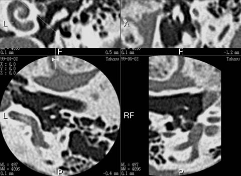
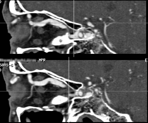
It has been a dream for radiologists to realize the "isotropic voxel" in CT where resolution is the same in x-, y- and z-axis since it will totally eliminate the partial volume averaging effect due to slice thickness. Half millimeter slices provides a true isotropic voxel in 24-cm field of view (FOV) and provides near-isotropic voxels in an 18-cm FOV (see Isotropic voxel). This means that the resolution of coronal and sagittal section is the same as that of an axial slice as shown in our clinical examples. Similar improvement can be observed in body-CT diagnosis with the use of 1-mm slices. I personally believe that this represents evolutional progress not only for CT diagnosis but also for all the slice-based diagnostic modalities.
As for the comparison of image characteristics of CT and MRI, it is believed that MRI is characterized by it's superb contrast resolution. However, it is a standard feature for current MRI units to provide slab thickness of 1-mm or less (e.g. MR angiography of the head using 3D-SPGR sequence, or CISS sequence for MR cisternography). Computed tomography, which is believed to be characterized by it's speed and superior spatial resolution, should be superior than MRI in terms of z-axis spatial resolution (slice thickness), otherwise CT examination may have little value in various clinical situations. This is especially true in the diagnosis of neuroradiological diseases, in which detailed interpretation of smaller anatomy is required.
For these reasons, I, as a neuroradiologist, cannot accept the concept of detector design in which the minimum detector aperture is 1.25-mm.
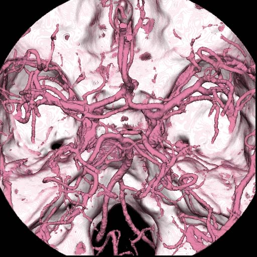
One of the serious shortcomings in thinner slice scanning is a limited scanning range - the thinner the slice is, the gerater is the scanning time required to cover the target organ(s). Two rows of 0.5-mm detector array will only works for the scanning of small organ such as middle ear. On the contrary, orbita, circle of Willis, facial bone, entire skull and brain or even the cervical and lumber spine are accessible to a 4-row system.
Another disadvantage of two 0.5-mm rows is that it is not possible to extract full advantages of pitch 3 scanning where the image quality is superior to other pitches (note that the pitch 3 scanning necessarily require 4 rows of detectors).
For these reasons, I disagree the concept of two rows of 0.5-mm slice.
Theoretically, a half-millimeter slice is effective in eliminating partial volume effect. However, it is extremely difficult to maintain sufficient image quality duaring actual scanning. Principal subjects for 0.5-mm scanning are as follows:
1. Geometrical accuracy
Since the detector aperture is extremely small compared with conventional CT, inaccuracy in scan geometry such as mechanical vibrations can cause errors in data measurement. This condition can be more serious when it is combined with 0.5-second rotation. One of the important factor is the scanning mechanism. The linear motor drive is far superior to a conventional belt-drive mechanism in reducing mechanical vibrations. Although it is costly, we believe that the linear motor drive is essential to maintain geometrical accuracy.
The second factor is the stability of the X-ray focal spot. The straddle bearing structure which is employed in the Aquilion contribute to a stable focal spot, eliminating these errors. Our tube posseses another advantage in geometrical accuracy, in which is achieved by the elimination of off-focal radiation by the employment of anode grounding structure (see X-ray tube).
2. Signal-to-noise ratio (s/n)
In a half-millimeter slice, the number of X-ray photons is 50% of a 1-mm slice. Moreover, 0.5-second rotation makes it half of 1-second scan, resulting 75% decrease in X-ray photons compared with conventional 1-mm / 1-second scan. Obviously, it is difficult to maintain good s/n in half-millimeter, half-second scanning.
The first step to maintain s/n is to increase X-ray output. This task is not easy because, for a half-millimeter slice, it is essential to employ a small focal spot size to minimize the blurring due to penumbra. A new X-ray system which consist of an oil-less, on-scanner generator and anode-grounded tube allows us to use 300-mA at 120-kV, or 260-mA at 135kV even with small focal spot.
Next mean is to secure sufficient detector aperture. In multislice detector, it is important to adjust the shape of neighboring detector edges. Smooth detector edges of Aquilion helps to eliminate tungsten shields to cover detector edges resulting successful maintain of wider detector aperture.
Increase in signal level can also be achieved by the proper adjustment of detector gain. Our detector system equipped with individual gain adjustment capability according to each detector aperture.
Elimination of noise is also an important factor. The gantly cover of Aquilion is electro-magnetically shielded in order to eliminate the noise from outside of the gantly.
-to be continued