

News & Topics
- June 14, 2021
- April 1, 2021
- March 14, 2021
Introduction
Members
Faculty members
Medical Technology
-
 Masato Abe
(Professor)
-
 Takamasa Yanagida
(Senior Assistant Professor) -
 Chiyuki Kaneko
(Senior Assistant Professor) -
 Kazuya Shiogama
(Senior Assistant Professor) -
 Masaya Hirayama
(Assistant)
Graduate student
Research Theme
Clinicopathological Diagnosis of Brain Tumors Indispensable for Patient’s Treatment
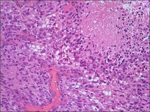 Histological findings of malignant brain tumor (glioblastoma)
1) Examination of technical methods of immunostaining
2) Identification of the origins of tumor cells
3) Check of proliferative potential
4) Evaluation of drug tolerance to chemotherapy
5) Investigation of genetic alteration
(Masato Abe)
Carcinogenic mechanism of NDMA-induced renal tumorigenesis in SD and F344 rats
(Takamasa Yanagida)
-
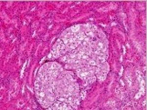 Renal cell carcinoma
-
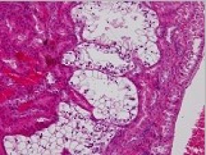 Adenoma
Differentiation of Malignant Mesothelioma from Adenocarcinoma Cells Appearing in Body Fluid -Using Immunohistology and Electron Microscopy
It can be difficult to differentiate malignant mesothelioma from adenocarcinoma cells or reactive mesothelial cells in body fluids. Therefore, we would like to examine the differentiation between adenocarcinoma cells and malignant mesothelioma cells appearing in body fluids using immunohistology, electron microscopy, and fluorescence in situ hybridization.
(Chiyuki Kaneko)
-
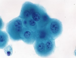 Malignant mesothelioma cells in pleural fluid
-
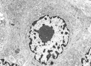 Electron Microscope Image of Malignant Mesothelioma<br />Nucleolus is clear
From the standpoint of pathology technologists, I have focused on developing "pathological techniques applicable to the routine work of pathological technologists.“ I specialize in improving pathological techniques, such as establishing highly sensitive in situ hybridization (Fig. 1) and ecological Grocott stain (Fig. 2). Currently, I am working on the following research project.
-
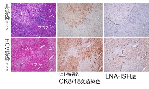 Figure 1: Highly sensitive in situ hybridization method for HCV detection
There are no good marketed antibodies against HCV and it is currently difficult to prove by immunostaining. A highly sensitive in situ hybridization method combining LNA-DNA oligoprobe and tyramide sensitization can detect HCV.
-
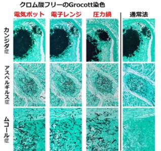 Figure 2: Environmentally friendly Grocott dyeing
The combination of heat treatment and periodic acid treatment enables the same level of dyeability as the conventional method without chromic acid.
1)Histochemistry of Neutrophil Extracellular Traps (NETs)
2)Morphological and functional evaluation of bacterial vaginosis using cytological specimens
3)Histochemistry of new cell death "necroptosis
-
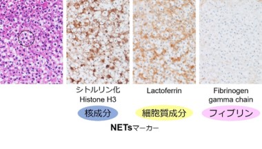 Figure 3-1: Differentiation between NETs and fibrin in HE-stained specimens (thin fibers).
Most of the fine fibers are positive for NETs markers and negative for fibrin markers, thus identifying them as NETs. (Acute appendicitis)
(Kazuya Shiogama) -
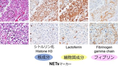 Figure 3-2: Differentiation between NETs and fibrin in HE-stained specimens (thick fibers).
Thick fibers indicate the coexistence of NETs and fibrin (NETs marker positive, fibrin marker positive) or fibrin (NETs marker negative, fibrin marker positive). (Upper row: Legionella pneumophila, Lower row: Liver abscess)
Organelles, represented by mitochondria, change their size and shape in response to the surrounding environment. Naturally, the cells formed by the myriad of variously shaped organelles each have their unique shape, and these cells form tissues and function as part of organs. To understand the pathogenesis of various diseases, we believe it is necessary to understand the complex hierarchical structure of living organisms in detail down to the nano-level, thus we are conducting ultrastructural analysis using the following morphological approaches.
- Seamless ultrastructure analysis by correlative light and electron microscopy
- Analysis of three-dimensional ultrastructure in paraffin sections by expansion microscopy
-
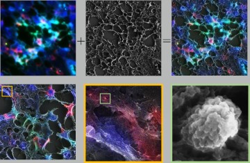 The workflow for optical microscopy to electron microscopy
When we take an electron microscope image of the same spot taken with an optical microscope, we can simultaneously prove the ultrastructure and expression.
-
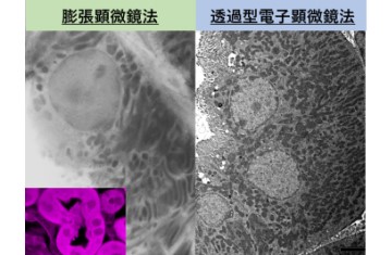 Nano-imaging of urinary tubules
Expansion microscopy method allows optical microscopes to have a high resolution close to electron microscope images.
Academic Activities
Manuscripts
2021
- Tsutsumi Y, Shiogama K, Sakurai K, Arase T, Domoto H: p16 immunostaining can avoid overdiagnosis in postmenopausal cervical cytology. IRJMMS. 9 (1): 1-8, 2021.
- Yamada S, Muto J, Iba S, Shiogama K, Tsuyuki Y, Satou Akira, Ohba S, Murayama K, Sugita Y, Nakamura S, Yokoo H, Tomita A, Hirose Y, Tsukamoto T, Abe M: Primary central nervous system lymphomas with massive intratumoral hemorrhage: clinical, radiological, pathological and molecular evaluation of six cases. Neuropathology. https://doi.org/10.1111/neup.12739, 2021.
- Shi H, Niimi A, Takeuchi T, Shiogama K, Mizutani Y, Kajino T, Inada K-I, Hase T, Hatta T, Shibata H, Fukui T, Chen‐Yoshikawa TF, Nagano K, Murate T, Kawamoto Y, Tomida S, Takahashi T, Suzuki M: CEBPγ facilitates lamellipodia formation and cancer cell migration through CERS6 upregulation. Cancer Sci. doi: 10.1111/cas.14928, 2021.
- Li, C., Onouchi, T., Hirayama, M., Sakai, K., Matsuda, S., Yamada, N. O., & Senda, T. (2021). Morphological and functional abnormalities of hippocampus in APC1638T/1638T mice. Medical molecular morphology, 54(1), 31–40. https://doi.org/10.1007/s00795-020-00257-3
- Yoshimitsu Y Katoh, Masaya Hirayama, Takanori Onouchi, Chiyuki Kaneko, Kazuyoshi Sakai, Masato Abe (2021). Necessity of glutaraldehyde-containing fixative to search for silver-positive bodies (nucleolus-like inclusion bodies) in the central nervous system cytoplasm. Journal of Analitical Bio-Science,44(3), 89-93.
2020
- Shigeo Ohba, MD, PhD, Kazuhiro Murayama, MD, PhD, Kiyonori Kuwahara, MD, Eriel Sandika Pareira, MD, Shunsuke Nakae, MD, PhD, Yuya Nishiyama, MD, PhD, Kazuhide Adachi, MD, PhD, Seiji Yamada, MD, PhD, Hikaru Sasaki, MD, PhD, Naoki Yamamoto, MD, PhD, Masato Abe, MD, PhD, Joydeep Mukherjee, PhD, Mitsuhiro Hasegawa, MD, PhD, Russell O Pieper, PhD, Yuichi Hirose, MD, PhD, The Correlation of Fluorescence of Protoporphyrinogen IX and Status of Isocitrate Dehydrogenase in Gliomas, Neurosurgery, Volume 87, Issue 2, August 2020, Pages 408–417, https://doi.org/10.1093/neuros/nyz524
- Takahashi, Y., Tsutsumi, Y., Takeuchi, C., Shiogama, K., Mizutani, Y., Inada, K. I., Yamamichi, N., & Koike, K. (2020). Nuclear staining of claudin-18 is a new immunohistochemical marker for diagnosing intramucosal well-differentiated gastric adenocarcinoma. Pathology international, 70(9), 644–652. https://doi.org/10.1111/pin.12978
- Hiramatsu, N., Yamamoto, N., Isogai, S., Onouchi, T., Hirayama, M., Maeda, S., Ina, T., Kondo, M., & Imaizumi, K. (2020). An analysis of monocytes and dendritic cells differentiated from human peripheral blood monocyte-derived induced pluripotent stem cells. Medical molecular morphology, 53(2), 63–72. https://doi.org/10.1007/s00795-019-00231-8
2019
- Ohba, S., Murayama, K., Nishiyama, Y., Adachi, K., Yamada, S., Abe, M., Hasegawa, M., & Hirose, Y. (2019). Clinical and Radiographic Features for Differentiating Solitary Fibrous Tumor/Hemangiopericytoma From Meningioma. World neurosurgery, 130, e383–e392. https://doi.org/10.1016/j.wneu.2019.06.094
- Kuwahara, K., Ohba, S., Nakae, S., Hattori, N., Pareira, E. S., Yamada, S., Sasaki, H., Abe, M., Hasegawa, M., & Hirose, Y. (2019). Clinical, histopathological, and molecular analyses of IDH-wild-type WHO grade II–III gliomas to establish genetic predictors of poor prognosis. Brain tumor pathology, 36(4), 135-143. https://doi.org/10.1007/s10014-019-00348-9
- Ohba, S., Yamada, Y., Murayama, K., Sandika, E., Sasaki, H., Yamada, S., Abe, M., Hasegawa, M., & Hirose, Y. (2019). c-Met Expression Is a Useful Marker for Prognosis Prediction in IDH-Mutant Lower-Grade Gliomas and IDH-Wildtype Glioblastomas. World neurosurgery, 126, e1042–e1049. https://doi.org/10.1016/j.wneu.2019.03.040
- Sakaguchi, Y., Yamamichi, N., Tomida, S., Takeuchi, C., Kageyama-Yahara, N., Takahashi, Y., Shiogama, K., Inada, K. I., Ichinose, M., Fujishiro, M., & Koike, K. (2019). Identification of marker genes and pathways specific to precancerous duodenal adenomas and early stage adenocarcinomas. Journal of gastroenterology, 54(2), 131–140. https://doi.org/10.1007/s00535-018-1489-4
- Okamura, S., Osaki, T., Nishimura, K., Ohsaki, H., Shintani, M., Matsuoka, H., Maeda, K., Shiogama, K., Itoh, T., & Kamoshida, S. (2019). Thymidine kinase-1/CD31 double immunostaining for identifying activated tumor vessels. Biotechnic & histochemistry : official publication of the Biological Stain Commission, 94(1), 60–64. https://doi.org/10.1080/10520295.2018.1499962
- Hamed, M. A., Sayed, R. H., Shiogama, K., Eltaher, M. A., Suzuki, K., & Nakata, S. (2019). Localisation of basic fibroblast growth factor in cholesteatoma matrix: an immunochemical study. The Journal of laryngology and otology, 133(3), 183–186. https://doi.org/10.1017/S0022215119000112
Conference
2021
- Masaya Hirayama, Takanori Onouchi, Kazuya Shiogama, Yoshimitsu Katoh, Kazuo Takahashi, Abe Masato Structural analysis of intracellular inclusion bodies localized in the paraventricular nucleus of the hypothalamus in normal mice The 53rd Annual Meeting of the Japanese Society for Clinical Molecular Morphology (to be announced in October)
2020
- Ayano Michiba, Kazuya Shiogama, Masaya Hirayama, Seiji Yamada, Takanori Onouchi, Chiyuki Kaneko, Takamasa Yanagida, Tetsuya Tsukamoto, Masato Abe Tumor-associated macrophages (TAMs) are involved in the formation of macrophage extracellular traps (METs) in glioblastoma The 109th Annual Meeting of the Japanese Society of Pathology
Book
- 水口國雄編集:染色法のすべて.医歯薬出版.p.47, 2021
- 「臨床検査学入門」編集委員会:医学領域における臨床検査学入門第4版.KTC中央出版.2018
- 標準理学療法学・作業療法学、病理学 第4版、医学書院、2017
Access
- Access to Fujita Health University ⇒ Fujita Health University Homepage
- Access to Morphology & Pathology 314, 3rd ⇒ University Building 3