
Animal Imaging Device
In vivo Imaging System
| Model (Manufacturer) |
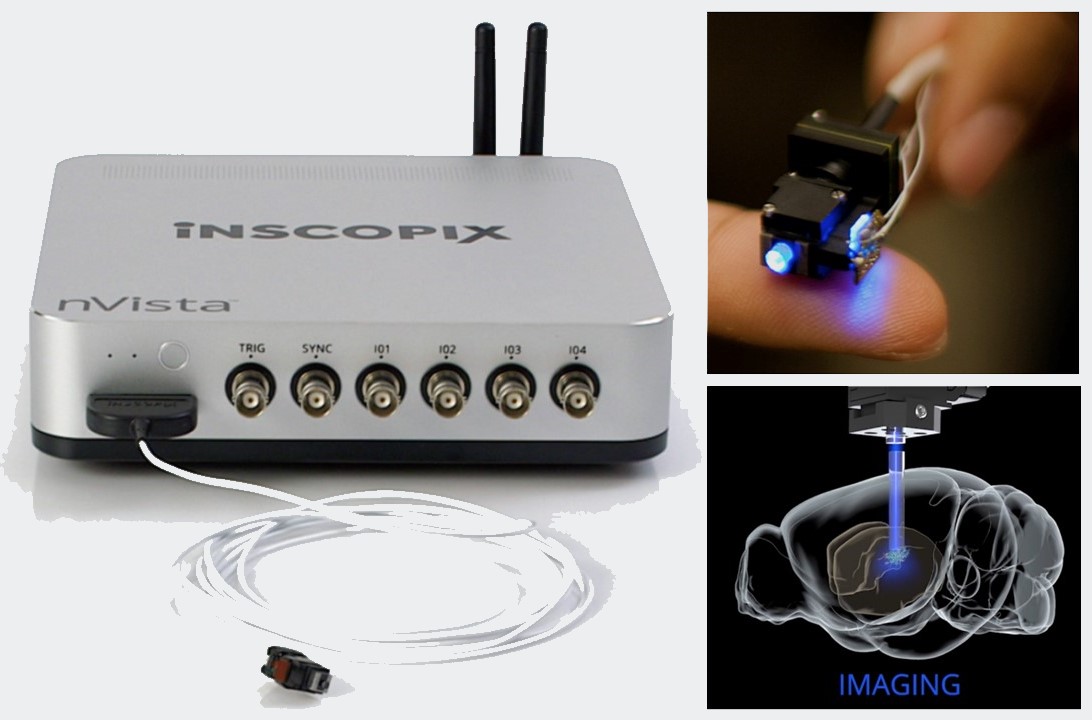
nVoke2.0 Integrated Imaging and Optogenetics System for Rodents (Inscopix) |
| Features and Specifications, etc. | - Integrates unique Ca2+ imaging technology and optogenetics to measure neural activity in real time while controlling the behavior of small animals. - Allows for simultaneous Ca 2+ imaging and light stimulation in small animals during behavior. - Capable of detecting hundreds of cells at the single-cell level. - Easy focus adjustment in the vertical direction. - Capable of synchronization with external devices through TTL control. 【Specifications】 System Style: Single-channel reflected fluorescence microscope Microscope light source: LED Lens aperture: NA 0.5 Field of view (FOV): 900 µm x 650 µm Field of view (pixels): 1280 x 800 pixels Focal length when using Proview lenses: 300 µm Effective focusing distance: 300 µm Z-axis resolution of focus adjustment: <0.5 µm Frame rate: 5-60 FPS Size: 8.8 mm x 15 mm x 22 mm Weight: 2.0 g Data cable: 2.5 m GPIO and digital IO: 8 digital GPI inputs 8 digital GPO outputs 4 analog GPIO I/O 1 SYNC output 1 TTL input 5 USB 3.0 ports Connection: WiFi connection, LAN cable Storage: 2TB SSD, SD card |
| Location | International Center for Brain Science (Room 601, University Building 1) |
| Remarks | - When using this equipment, please ensure to reserve the device through the Joint Usage Research Facility Reservation System, as well as booking the laboratory at the Disease Model Education and Research Support Center. - When using the equipment, please be sure to fill out the logbook provided with it. - Please prepare consumables such as Proview lenses or normal lenses by yourself. |
| Equipment Manager Extension Number |
takahashi ・mizuguchi(Bioimaging Analysis Laboratory) 2645 |
| Usage Fee | 200 yen/hour |
機器写真
In vivo Bioluminescence, Fluorescence, and CT Imaging System
| Model (Manufacturer) |
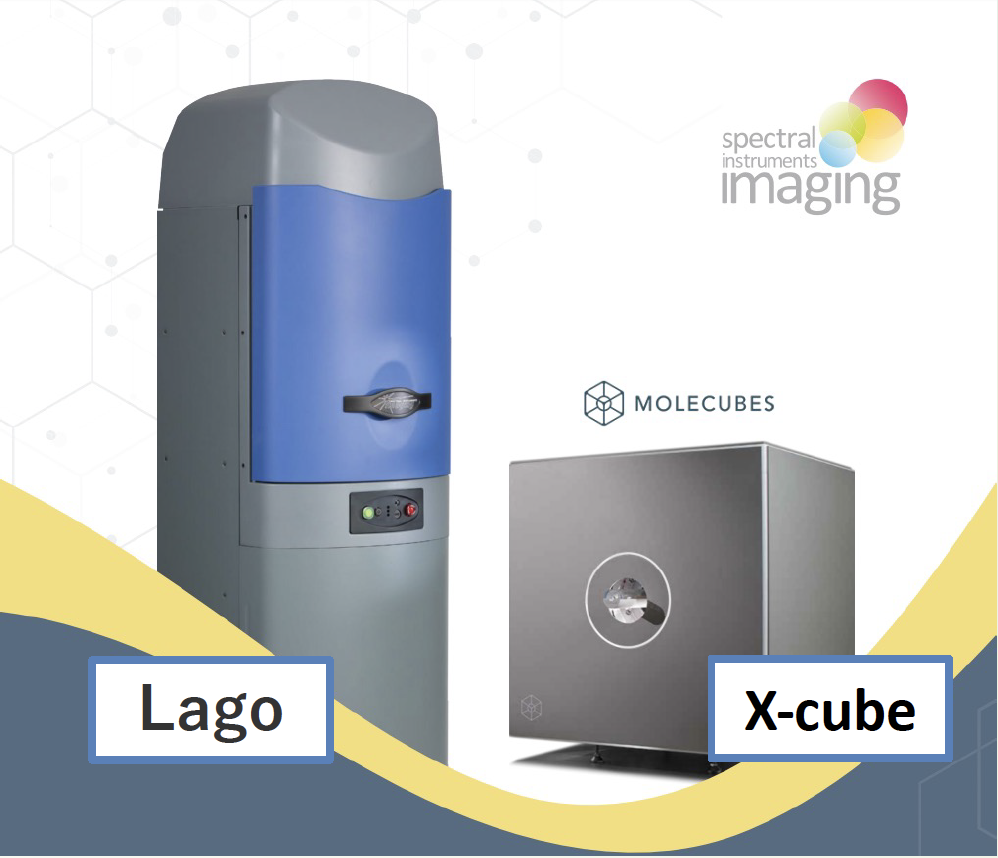
- In vivo bioluminescence and fluorescence imaging system Lago X - CT system Molecubes X-CUBE from Spectral Instruments Imaging (Lago-X) Molecubes (X-Cube) |
| Features and Specifications, etc. | - Features of the in vivo bioluminescence and fluorescence imaging system Lago Achieves high-sensitivity light imaging with an air-cooled CCD camera at -90℃. Achieves reduced autofluorescence and system noise through a patented high-brightness LED excitation method. Allows simultaneous measurement of up to ten mice in a wide imaging field during light imaging. Equipped with X-ray 2D imaging capabilities. Provides image analysis software free of charge. Camera cooling time from power on to measurement start is just under 5 minutes. - Features of the CT multimodality solution Molecubes' microCT device X-CUBE, inviCRO's VivoQuant analysis software, Utilizes In vivo ANALYTICS' quantitative 3D optical imaging solution to: estimate the three-dimensional spatial information and signal intensity of bioluminescence (BLI) and fluorescence (FLI) signals originating from within the body, allowing for overlay with high-resolution microCT. |
| Location | Disease Model Center (Conventional Multipurpose Room 3, B3 Floor, University Building 1) |
| Remarks | Disease Model Center (Conventional Multipurpose Room 3, B3 Floor, University Building 1) Remarks - Please ensure to book this equipment through the reservation system when using it. - When using the equipment, please be sure to fill out the logbook provided with it. - Please also reserve "Conventional Multipurpose Room 3" through the Disease Model Center's reservation site. - For inquiries related to animals, please consult the Disease Model Center. |
| Equipment Manager Extension Number |
takahashi ・mizuguchi(Bioimaging Analysis Laboratory) 2645 |
| Usage Fee | 500 yen/10 min. |
機器写真