
Confocal Microscopy
Confocal Laser Microscope
| Model (Manufacturer) |
LSM-710 (Carl Zeiss) |
| Features and Specifications, etc. |
Overview/Features An apparatus that utilizes the characteristics of confocal optical systems to obtain optical cross-sectional images of samples. Since only information from the in-focus plane is utilized, clear images without flare can be obtained. Taking Z-stack images to construct a three-dimensional image. It is equipped with a culture device, enabling live imaging. Specifications Inverted Microscope (Axio Observer Z1) Laser: 405nm, Argon (458/488/514nm), HeNe (543/561/594/633nm) Objective Lenses: 5X, 10X, 20X, 40X(oil), 63X(oil) Culture Equipment (35mm glass bottom dish) |
| Location | Bioimaging Analysis Laboratory(Electron Microscope Room, Room 115, University Building 1) |
| Remarks | - Under the supervision of faculty, undergraduate students are generally only allowed to observe. - Please be sure to apply for eligibility through the equipment reservation system and book your reservation after approval. - Training within the course or similar settings is prohibited. - Sensing the risk of infection, bringing in samples is prohibited. |
| Equipment Manager Extension Number |
Mizuguchi (Bioimaging Analysis Laboratory) 2645 |
| Usage Fee | 300 yen/hour |
Click here for equipment manual
Equipment Photo

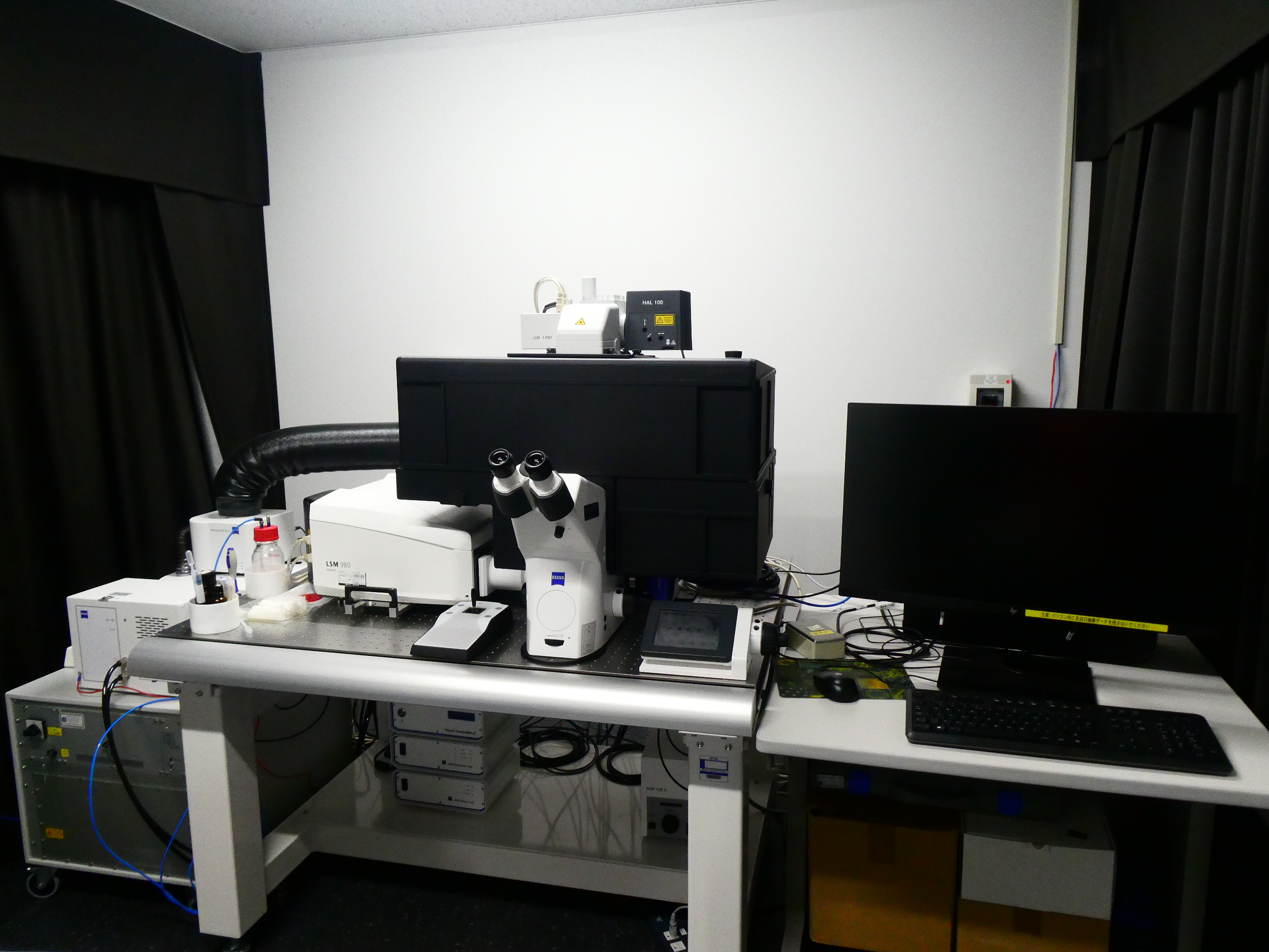
Confocal Laser Microscope
| Model (Manufacturer) |
LSM-980 (Carl Zeiss) |
| Features and Specifications, etc. |
Overview/Features In addition to the confocal detector, it is equipped with an Airyscan detector, enabling high-sensitivity and low-damage super-resolution imaging. Thanks to its low phototoxicity and high-speed capabilities, it allows for longer periods of live imaging compared to LSM710. Specifications Inverted Microscope (Axio Observer 7) Laser: 405/445/488/514/561/639nm Objective Lenses: 2.5X, 10X, 20X, 25X(autocorr), 40X(oil) ,63X(oil) Incubator (35mm glass bottom dish) |
| Location | Bioimaging Analysis Laboratory(Electron Microscope Room, Room 115, University Building 1) |
| Remarks | - Under the supervision of faculty, undergraduate students are generally only allowed to observe. - Please be sure to apply for eligibility through the equipment reservation system and book your reservation after approval. - Training within the course or similar settings is prohibited. - Sensing the risk of infection, bringing in samples is prohibited. |
| Equipment Manager Extension Number |
Mizuguchi (Bioimaging Analysis Laboratory) 2645 |
| Usage Fee | 900 yen/hour |
Click here for equipment manual
Equipment Photo
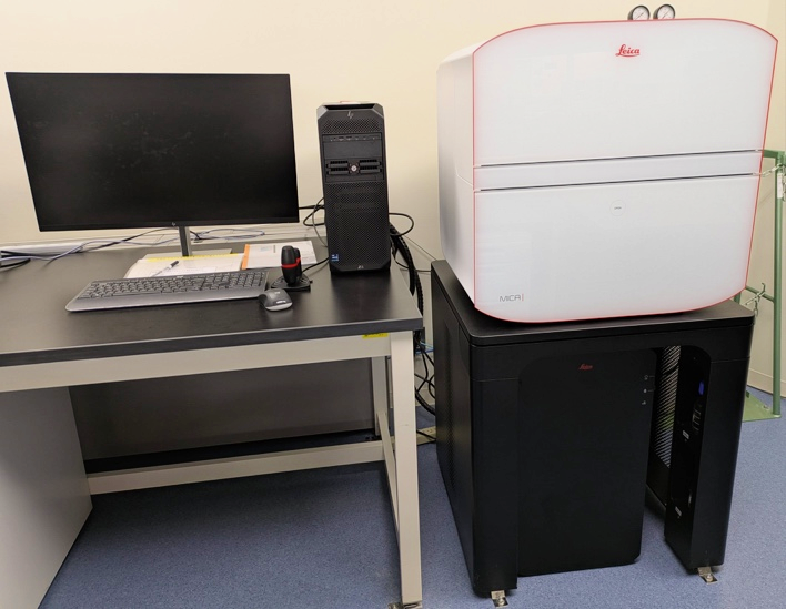
Imaging Microhub
| Model (Manufacturer) |
MICA (Leica) |
| Features and Specifications, etc. | Overview/Features - Integrated microscope eliminates the need for a darkroom and automatically sets the imaging conditions in confocal to save a significant amount of time. - Four detectors for Confocal and four color cameras for WideField are equipped, enabling simultaneous imaging of up to four fluorescent colors. - The sample that can be observed is a multi-well plate, a slide (up to 4 slides can be mounted), a chamber slide, or a 35mm dish. Specifications Inverted microscope Excitation wavelength: Camera mode (LED:365/470/555/625nm) Confocal mode (diode laser:405/488/561/638nm) Detector: Camera mode: 5 million pixels, CMOS camera (with color mode), 4 channels Confocal mode: Hyd FS, 4 channels Detection wavelength range: Camera mode: 420nm-705nm Confocal mode: 415nm-750nm Objective lenses: 1.6X, 10X, 20X, 63X (auto immersion) Z-stack Thunder function Lightning function |
| Location | Central Research Center (Room 317, University Building 1) |
| Remarks | - Please be sure to apply for eligibility through the equipment reservation system and book your reservation after approval. - To take out data, please use a USB or similar device that has been virus-checked by PC for taking out data at the Central Center. |
| Equipment Manager Extension Number |
Mizuguchi (Bioimaging Analysis Laboratory) 2645 |
| Usage Fee | 300 yen/hour |
Click here for equipment manual
Equipment Photo
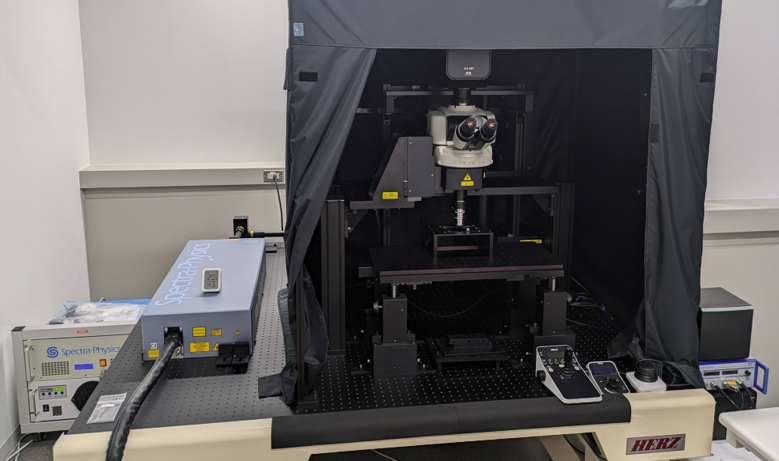
Multiphoton Excitation Microscopy
| Model (Manufacturer) |
AX-RMP(NIKON) |
| Features and Specifications, etc. |
Overview/Features Multiphoton excitation microscopes use two-photon excitation and are capable of acquiring clearer images of molecular dynamics and microstructures deeper in vivo, with less fading and photo-damage than conventional confocal microscopes. It is effective for in vivo imaging in physiological studies of the cranial nervous system, cardiovascular system, digestive system, respiratory system, and pathophysiological studies of cancer and neuropsychiatric diseases. Specifications Excitation wavelength: 700nm-1040nm variable femtosecond pulse laser Detector: NDD ch1-ch4: GaAsP Detection wavelength range: 400nm-650nm Maximum resolution: 8192 X 8192 (Galvano scanner) Fastest frame rate: 240fps (512 X 16) Galvano 720fps (2048 X 16) Resonant Upright microscope Motorized XY stage (XY movement range 65 X 65mm) Objective piezo unit (movement range 400㎛) Objective lenses: 10X, 16X(water immersion), 20X(water immersion), 25X(water immersion), 40X(water immersion) |
| Location | Open Lab(Room 605, University Building 1) |
| Remarks |
- Use in compliance with the rules of use. - Please be sure to apply for eligibility through the equipment reservation system and book your reservation after approval. |
| Equipment Manager Extension Number |
Yamashita(PhysiologyⅡ)2465 Mizuguchi (Bioimaging Analysis Laboratory)2645 |
| Usage Fee | 1,000 yen/hour |
Equipment Photo