
Electron Microscopes
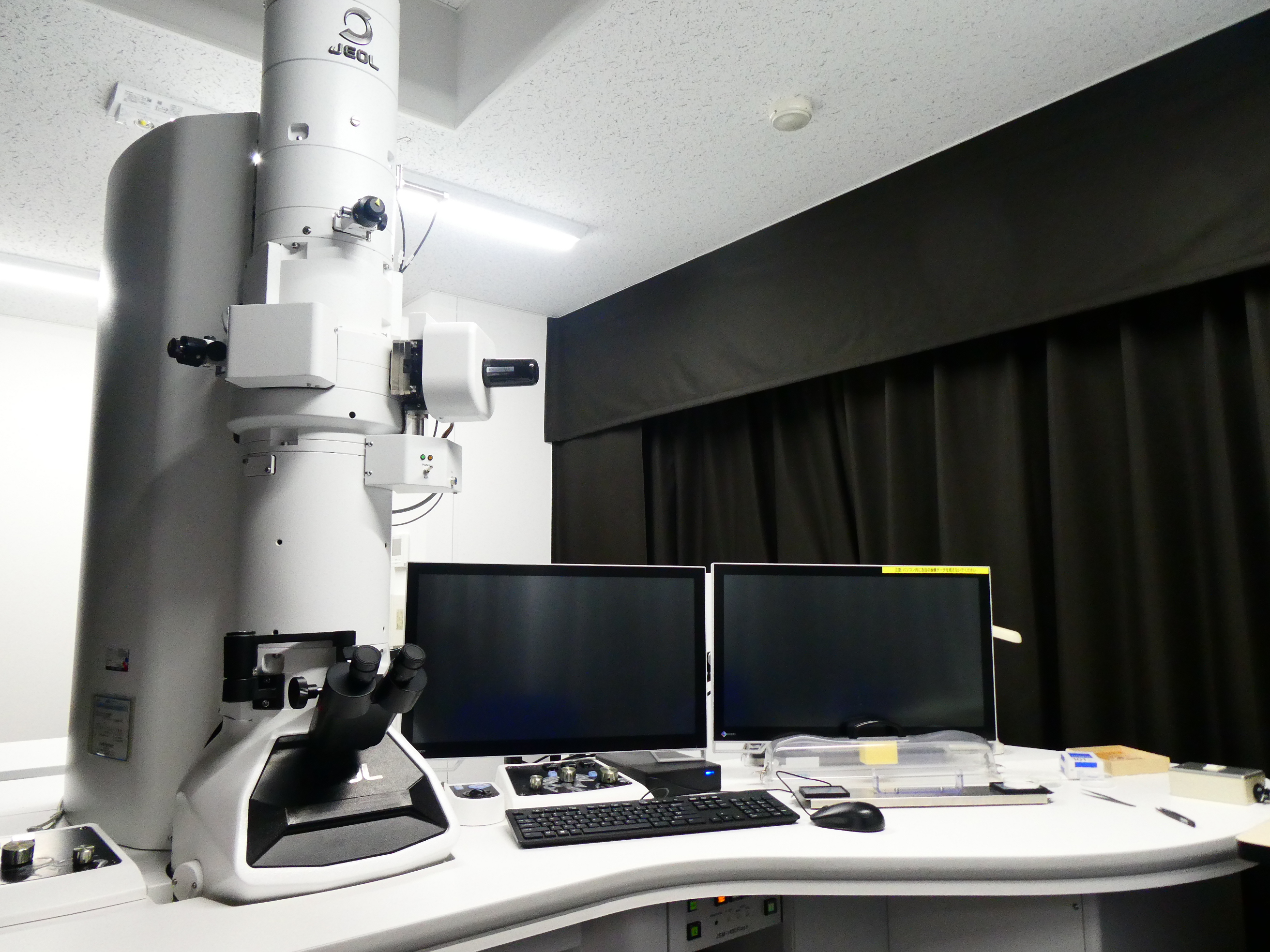
Transmission Electron Microscope
| Model (Manufacturer) |
JEM-1400Flash (JEOL) |
| Features and Specifications, etc. | Overview/Features Device used to observe cross sections of small samples that cannot be seen with an optical microscope. Electrons are irradiated onto ultrathin sections less than 100nm thick, allowing observation of the fine structure of the sample by imaging the electrons that pass through the ultrathin sections. Specifications TEM resolution: 0.2nm, acceleration voltage: 10-120kV, magnification: x 10 to 1,200,000, maximum sample tilt angle (Tilt-X), exhaust system: TMP, equipped with JEOL manufactured sCMOS camera, ultra-wide field montage system Limitless Panorama (LLP) equipped, optical microscope image link feature Picture Overlay equipped, etc. |
| Location | Bioimaging Analysis Laboratory (Electron Microscope Room, Room 115, University Building 1) |
| Remarks | - Under the supervision of faculty, undergraduate students are generally only allowed to observe. - When using this equipment, always receive handling instructions from the person in charge before using it for the first time. - Please ensure to book this equipment through the reservation system when using it. |
| Equipment Manager Extension Number |
Tominaga (Bioimaging Analysis Laboratory) 2320 |
| Usage Fee | 300 yen/hour |
Equipment Photo
Scanning Electron Microscope
| Model (Manufacturer) |
JSM-7610FPlus (JEOL) |
| Features and Specifications, etc. | Overview/Features Device used for observing the surfaces of small objects that cannot be seen with an optical microscope.When an electron beam emitted from a field emission electron gun is directed at a sample, the information from the electrons emitted from the sample can be used to observe the sample's surface topography and composition. Specifications Resolution: 0.8 nm (at 15 kV acceleration voltage), 1.0 nm (at 1 kV GB mode), 0.8 nm (at 1 kV GBSH mode), Analysis mode 3.0 nm (at 15 kV acceleration voltage, WD8 mm, emission current 5 nA), Acceleration voltage: 0.1 - 30kV, Magnification: Photographic magnification: x 25 to 1,000,000 (display at 120mm x 90mm), Display magnification: x 75 to 3,000,000 (display at 1,280 x 960 pixels), Electron gun: In-lens Schottky field emission electron gun, Lens system: Condenser lens (CL), Aperture angle optimization lens (ACL), Semi-in-lens objective lens (OL), Sample stage: Full eucentric goniometer stage, 5-axis motor-driven, Exhaust system: Electron gun chamber/intermediate chamber ultra-high vacuum dry exhaust system by ion pump, Sample chamber dry exhaust system TMP exhaust system, etc. |
| Location | Bioimaging Analysis Laboratory (Electron Microscope Room, Room 115, University Building 1) |
| Remarks | - Under the supervision of faculty, undergraduate students are generally only allowed to observe. - When using this equipment, always receive handling instructions from the person in charge before using it for the first time. - Please ensure to book this equipment through the reservation system when using it. |
| Equipment Manager Extension Number |
Tominaga (Bioimaging Analysis Laboratory) 2320 |
| Usage Fee | 300 yen/hour |
Equipment Photo

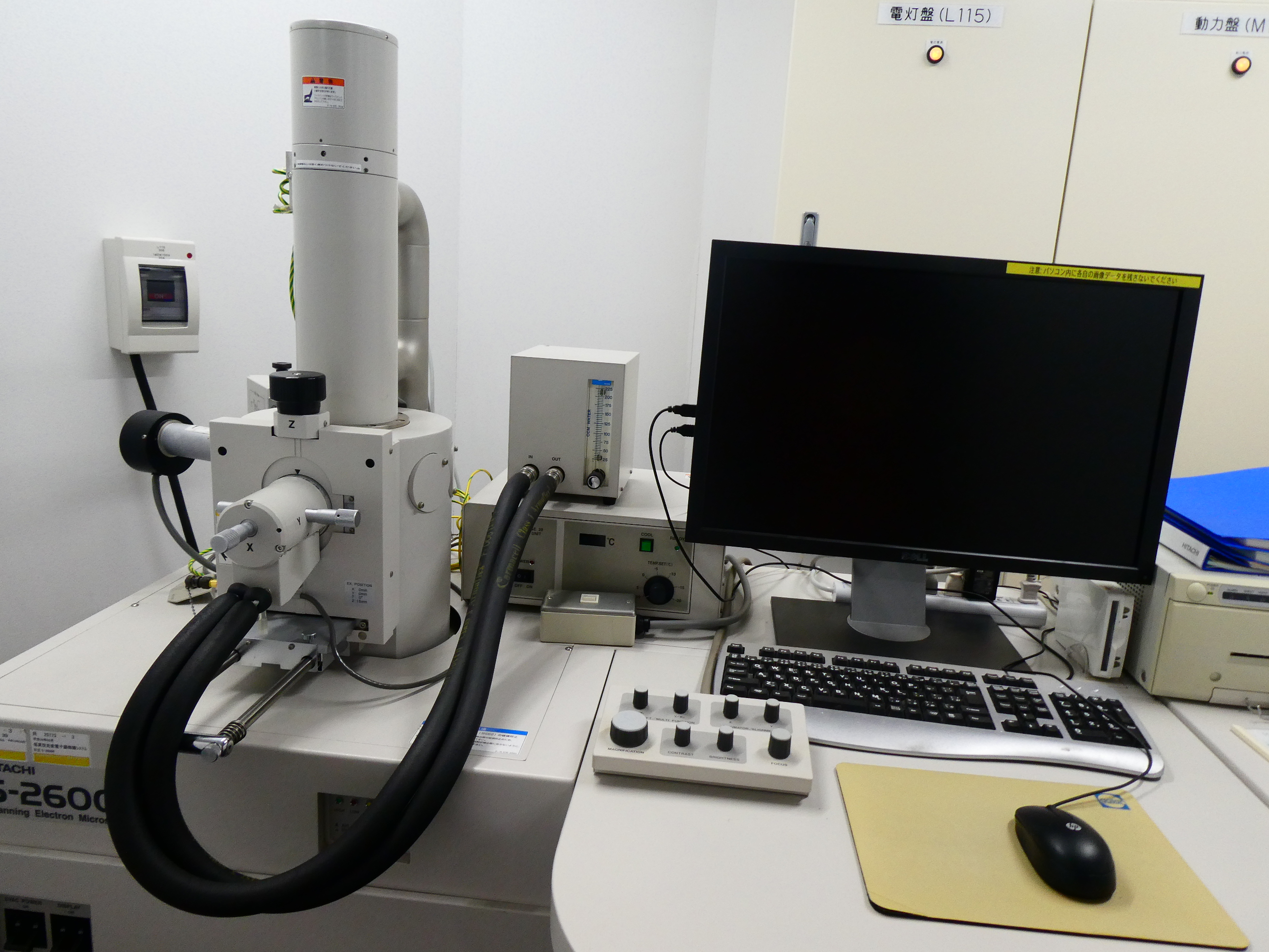
Scanning Electron Microscope
| Model (Manufacturer) |
HITACHI S-2600 (HITACHI) |
| Features and Specifications, etc. | Overview/Features Device used for observing the surfaces of small objects that cannot be seen with an optical microscope. When an electron beam emitted from a tungsten filament electron gun is directed at a sample, the information from the electrons emitted from the sample can be used to observe the sample's surface topography and composition. Specifications Resolution: Secondary electron image resolution 4 nm (high vacuum mode), backscattered electron image resolution 5 nm (low vacuum mode), Acceleration voltage: 0.5–30 kV, 0.5–5 kV, 5–30 kV, Magnification: x 15 to 300,000, etc. |
| Location | Bioimaging Analysis Laboratory (Electron Microscope Room, Room 115, University Building 1) |
| Remarks | - When using this equipment, always receive handling instructions from the person in charge before using it for the first time. - Please ensure to book this equipment through the reservation system when using it. |
| Equipment Manager Extension Number |
Tominaga (Bioimaging Analysis Laboratory) 2320 |
| Usage Fee | - |
Equipment Photo