

Electron Microscope Sample Preparation Equipment
Ultramicrotome
| Model (Manufacturer) |
Reichert Ultracut H (Reichert) |
| Features and Specifications, etc. | Overview/Features Device used to create ultrathin sections for electron microscopy observation. Using glass knives or diamond knives, it is possible to produce semi-ultrathin sections (thickness between 500nm and 1μm) and ultrathin sections (thickness less than 100nm). |
| Location | Bioimaging Analysis Laboratory (Control Room, Room 113, University Building 1) |
| Remarks | - When using this equipment, always receive handling instructions from the person in charge before using it for the first time. - Please ensure to book this equipment through the reservation system when using it. |
| Equipment Manager Extension Number |
Tominaga (Bioimaging Analysis Laboratory) 2320 |
| Usage Fee | 100 yen/hour |
Equipment Photo

Ultramicrotome
| Model (Manufacturer) |
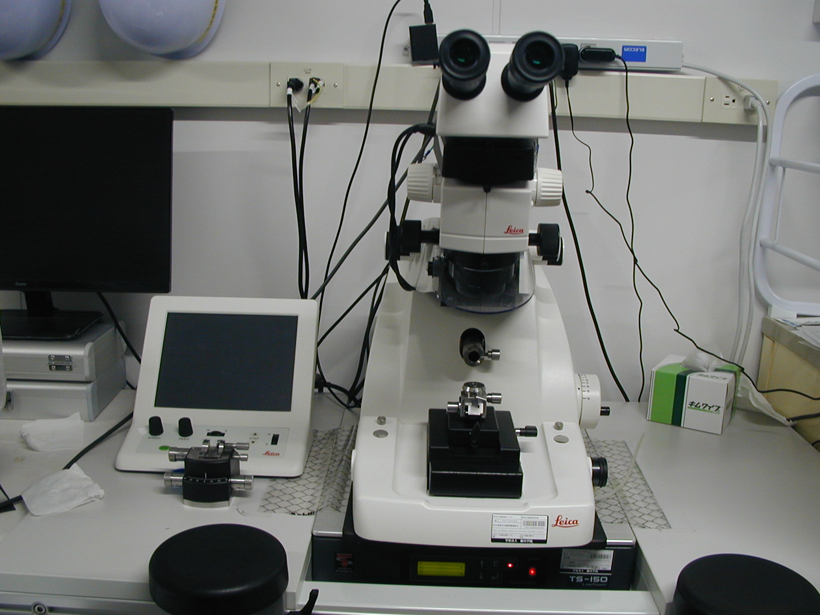
Leica EM UC7 (with anti-vibration tables) (Leica) |
| Features and Specifications, etc. | Overview/Features Device used to create ultrathin sections for electron microscopy observation.Using glass knives or diamond knives, it is possible to produce semi-ultrathin sections (thickness between 500nm and 1μm) and ultrathin sections (thickness less than 100nm). By installing additional anti-vibration tables, it is possible to prevent variations in thickness (chatter) caused by vibrations during the production of ultrathin sections. |
| Location | Bioimaging Analysis Laboratory (Control Room, Room 113, University Building 1 ) |
| Remarks | - When using this equipment, always receive handling instructions from the person in charge before using it for the first time. - Please ensure to book this equipment through the reservation system when using it. |
| Equipment Manager Extension Number |
Tominaga (Bioimaging Analysis Laboratory) 2320 |
| Usage Fee | 100 yen/hour |
Equipment Photo
Ultramicrotome
| Model (Manufacturer) |
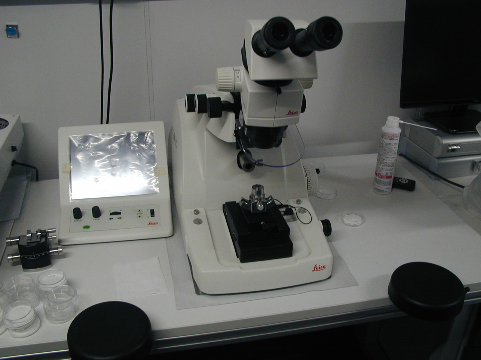
Leica EM UC7 (without anti-vibration tables) (Leica) |
| Features and Specifications, etc. | Overview/Features Device used to create ultrathin sections for electron microscopy observation.Overview/Features Device used to create ultrathin sections for electron microscopy observation. Using glass knives or diamond knives, it is possible to produce semi-ultrathin sections (thickness between 500nm and 1μm) and ultrathin sections (thickness less than 100nm). |
| Location | Bioimaging Analysis Laboratory (Control Room, Room 113, University Building 1) |
| Remarks | - When using this equipment, always receive handling instructions from the person in charge before using it for the first time. - Please ensure to book this equipment through the reservation system when using it. |
| Equipment Manager Extension Number |
Tominaga (Bioimaging Analysis Laboratory) 2320 |
| Usage Fee | 100 yen/hour |
Equipment Photo
Hydrophilic Treatment Equipment
| Model (Manufacturer) |
HDT-400 (JEOL) |
| Features and Specifications, etc. | Overview/Features Device for hydrophilizing hydrophobic transmission electron microscopy grids (mesh) and diamond knives. |
| Location | Bioimaging Analysis Laboratory (Control Room, Room 113, University Building 1) |
| Remarks | - When using this equipment, always receive handling instructions from the person in charge before using it for the first time. - Please ensure to book this equipment through the reservation system when using it. |
| Equipment Manager Extension Number |
Tominaga (Bioimaging Analysis Laboratory) 2320 |
| Usage Fee | - |
Equipment Photo

Glass Knife Maker
| Model (Manufacturer) |
EM KMR3 (Leica) |
| Features and Specifications, etc. | Overview/Features Device for making glass knives. Glass knives are used to produce semi-ultrathin sections (thickness between 500nm and 1μm). Specifications Operating principle: Balance breaking method, Score marks: Two types (preset), Scoring angle: 45°, Rod glass: For ultramicrotomes, freeze ultramicrotomes, and special glass for histological examinations, Dimensions: Length: 400mm, Width: 25.4mm, Thickness: 6.4mm, 8mm, 10mm, capable of being broken. Scoring wheel: tungsten carbide |
| Location | Bioimaging Analysis Laboratory (Control Room, Room 113, University Building 1 ) |
| Remarks | - When using this equipment, always receive handling instructions from the person in charge before using it for the first time. - Please ensure to book this equipment through the reservation system when using it. |
| Equipment Manager Extension Number |
Tominaga (Bioimaging Analysis Laboratory) 2320 |
| Usage Fee | - |
Equipment Photo

Vacuum Evaporation Device
| Model (Manufacturer) |
JEE-420T (JEOL) |
| Features and Specifications, etc. | Overview/Features Device for producing carbon support films suitable for electron microscopy observation. |
| Location | Bioimaging Analysis Laboratory (Electron Microscope Room, Room 115, University Building 1) |
| Remarks | - Please ensure to book this equipment through the reservation system when using it. |
| Equipment Manager Extension Number |
Tominaga (Bioimaging Analysis Laboratory) 2320 |
| Usage Fee | - |
Equipment Photo

Ion Sputtering Apparatus
| Model (Manufacturer) |
JFC-1500 (JEOL) |
| Features and Specifications, etc. | Overview/Features Device for conducting conductive treatment by coating the surface of non-conductive samples, such as biological specimens, with various metals, and for etching the surface of samples. Specifications Method: Bipolar opposing electrode discharge method, Mode: DC/AC coating, DC/AC etching, Coating target: Standard 50mm diameter Au (gold), etc. |
| Location | Bioimaging Analysis Laboratory (Electron Microscope Room, Room 115, University Building 1) |
| Remarks | - When using this equipment, always receive handling instructions from the person in charge before using it for the first time. - Please ensure to book this equipment through the reservation system when using it. |
| Equipment Manager Extension Number |
Tominaga(Bioimaging Analysis Laboratory) 2320 |
| Usage Fee | - |
Equipment Photo

Auto Fine Coater
| Model (Manufacturer) |
JEC-3000FC (JEOL) |
| Features and Specifications, etc. | Overview/Features Device for performing conductive treatment by coating the surface of non-conductive samples, such as biological specimens, with various metals. |
| Location | Bioimaging Analysis Laboratory (Electron Microscope Room, Room 115, University Building 1) |
| Remarks | - When using this equipment, always receive handling instructions from the person in charge before using it for the first time. - Please ensure to book this equipment through the reservation system when using it. |
| Equipment Manager Extension Number |
Tominaga (Bioimaging Analysis Laboratory) 2320 |
| Usage Fee | - |
Equipment Photo

Osmium Coater
| Model (Manufacturer) |
HPC-30 (Vacuum Device) |
| Features and Specifications, etc. | Overview/Features Device for performing conductive treatment by coating the surface of non-conductive samples, such as biological specimens, with osmium. |
| Location | Bioimaging Analysis Laboratory (Electron Microscope Room, Room 115, University Building 1) |
| Remarks | - Please ensure to book this equipment through the reservation system when using it. |
| Equipment Manager Extension Number |
Tominaga (Bioimaging Analysis Laboratory) 2320 |
| Usage Fee | - |
Equipment Photo

Ultraviolet Curing Device
| Model (Manufacturer) |
TUV-200 (DOSAKA EM) |
| Features and Specifications, etc. | Overview/Features Device used for creating water-soluble resin blocks used in techniques such as immunoelectron microscopy. Ultraviolet light is irradiated onto water-soluble resin to induce polymerization and hardening (UV polymerization). It is possible to fill the sample with N2 (nitrogen gas). The control and irradiation sections are separated, allowing for operation outside of the freezer. Sample capsule capacity is up to 60 capsules. UV irradiation can be performed at temperatures ranging from -70°C to room temperature. |
| Location | Bioimaging Analysis Laboratory (Electron Microscope Room, Room 115, University Building 1 ) |
| Remarks | - Please ensure to book this equipment through the reservation system when using it. |
| Equipment Manager Extension Number |
Tominaga (Bioimaging Analysis Laboratory) 2320 |
| Usage Fee | - |
Equipment Photo

Freeze Drying Equipment
| Model (Manufacturer) |
VFD-21S (Vacuum Device) |
| Features and Specifications, etc. | Overview/Features Device for drying biological samples for scanning electron microscopy. The water content is replaced by t-butyl alcohol, frozen, and then the t-butyl alcohol is sublimated in a vacuum. |
| Location | Bioimaging Analysis Laboratory (Electron Microscope Room, Room 115, University Building 1 ) |
| Remarks | - Please ensure to book this equipment through the reservation system when using it. |
| Equipment Manager Extension Number |
Tominaga (Bioimaging Analysis Laboratory) 2320 |
| Usage Fee | 100 yen/time |
Equipment Photo