

Optical Microscopes
Contents on page
- Bright Field Upright Microscope Camera System
- Bright Field Stereo Microscope Camera System
- Fluorescence Upright Microscope Camera System
- Fluorescence Stereo Microscope Camera System
- Fluorescence Inverted Microscope Camera System
- Discussion Upright Microscopes
- All-in-one Fluorescence Microscope System
- All-in-one Fluorescence Microscope System
- Digital Imaging System
- Multiplex Spatial analysis system
- Virtual Slide Cinematography Equipment
- Fluorescence & Bright Field Slide Scanners
- High Content Imaging System
- Analysis PC for ImageXpress data
- High Content Imaging System
- Opera Phenix Analysis PC
- Columbus Image Analysis Processing Software
Bright Field Upright Microscope Camera System
| Model (Manufacturer) |
BX-43, DP-70(Olympus) |
| Features and Specifications, etc. |
Overview/Features This is an inspection microscope suitable for brightfield observation. It is equipped with a CCD camera, enabling image capture on a PC. Specifications Upright Microscope Objective Lenses: 4X, 10X, 20X, 40X, 100X(oil) CCD Color Camera (DP70) |
| Location | Central Research Center (Room 317, University Building 1) |
| Remarks | - Please ensure to book this equipment through the reservation system when using it. |
| Equipment Manager Extension Number |
Mizuguchi (Bioimaging Analysis Laboratory) 2645 |
| Usage Fee | - |
Equipment Photo

Bright Field Stereo Microscope Camera System
| Model (Manufacturer) |
SZ-9, DP-71 (Olympus) |
| Features and Specifications, etc. |
Overview/Features A versatile stereo microscope that faithfully reproduces natural colors and shapes. Observation is conducted using brightfield and oblique illumination. It is equipped with a CCD camera, enabling image capture on a PC. Specifications Stereomicroscope Objective Lens: 1X Zoom 4.7-42.8 CCD Color Camera (DP71) |
| Location | Central Research Center (Room 317, University Building 1) |
| Remarks | - Please ensure to book this equipment through the reservation system when using it. |
| Equipment Manager Extension Number |
Mizuguchi (Bioimaging Analysis Laboratory) 2645 |
| Usage Fee | - |
Equipment Photo

Fluorescence Upright Microscope Camera System
| Model (Manufacturer) |
BX-51, DP-74 (Olympus) |
| Features and Specifications, etc. |
Overview/Features A upright microscope capable of high-resolution fluorescence imaging and excellent brightfield observation with vibrant color reproduction. It is equipped with a CCD camera, enabling image capture on a PC. Specifications Orthostatic Fluorescence Microscope Differential Interference Objective Lenses: 4X, 10X, 20X, 40X, 100X(oil) Fluorescence Filters: DAPI,GFP,YFP,TRITC CCD Color Camera (DP74) |
| Location | Central Research Center (Room 317, University Building 1) |
| Remarks | - Please ensure to book this equipment through the reservation system when using it. |
| Equipment Manager Extension Number |
Mizuguchi (Bioimaging Analysis Laboratory) 2645 |
| Usage Fee | - |
Equipment Photo

Fluorescence Stereo Microscope Camera System
| Model (Manufacturer) |
MZ-16F (Leica) |
| Features and Specifications, etc. |
Overview/Features Rotating the side knob on the transmitted light base enables both brightfield and darkfield observations. It is equipped with a mercury lamp, allowing for fluorescence observation. It is equipped with a CCD camera, enabling image capture on a PC. Specifications Stereo Fluorescence Microscope Objective Lenses: 1X, 5X Zoom 0.71-11.5 CCD Color Camera (DFC300FX) |
| Location | Central Research Center (Room 317, University Building 1) |
| Remarks | - Please ensure to book this equipment through the reservation system when using it. |
| Equipment Manager Extension Number |
Mizuguchi (Bioimaging Analysis Laboratory) 2645 |
| Usage Fee | - |
Equipment Photo

Fluorescence Inverted Microscope Camera System
| Model (Manufacturer) |
Axio Observer (Carl Zeiss) |
| Features and Specifications, etc. |
Overview/Features An inverted microscope capable of high-resolution fluorescence imaging. It is equipped with a CCD camera, enabling image capture on a PC. Specifications Inverted Fluorescence Microscope Differential Interference Objective Lenses: 5X, 10X, 20X, 40X, 63X(oil) Fluorescence Filters: DAPI,GFP,TRITC CCD Monochrome Camera (AxioCam MRm) |
| Location | Central Research Center (Room 317, University Building 1) |
| Remarks | - Please ensure to book this equipment through the reservation system when using it. |
| Equipment Manager Extension Number |
Mizuguchi (Bioimaging Analysis Laboratory) 2645 |
| Usage Fee | - |
Equipment Photo

Discussion Upright Microscopes
| Model (Manufacturer) |
Axio Imager2 (Carl Zeiss) |
| Features and Specifications, etc. |
Overview/Features Four eyepiece lenses are installed in addition to the main unit, but only three can be observed at the same time due to the installation location. Equipped with a CCD camera, so images can be acquired on a PC. Specifications Upright Microscope Objective Lenses: 1X, 2.5X, 5X, 10X, 20X, 40X, 100X(oil) Camera: CCD color camera(AxioCam MRc5) |
| Location | Central Research Center (Room 317, University Building 1) |
| Remarks | - Please ensure to book this equipment through the reservation system when using it. |
| Equipment Manager Extension Number |
Mizuguchi (Bioimaging Analysis Laboratory) 2645 |
| Usage Fee | - |
Equipment Photo

All-in-one Fluorescence Microscope System
| Model (Manufacturer) |
BZ-9000 (Keyence) |
| Features and Specifications, etc. |
Overview/Features Since the microscope is housed in a box, a darkroom is not necessary even during fluorescence observation. There are no eyepiece lenses, and the images are displayed on a PC screen. By changing the stage adapter, observation of slides, 35mm dishes, and multi-well plates is possible. Specifications Inverted Fluorescence Microscope Phase Contrast Objective Lenses: 4X, 10X, 20X, 40X(Phase Contrast), 100X(oil), 20X(Long Working Distance Phase Contrast) Fluorescence Filters: DAPI,GFP,TRITC Camera: CCD monochrome & color Z-stacking, tiling, multi-point shooting |
| Location | Central Research Center (Room 317, University Building 1) |
| Remarks | - Please ensure to book this equipment through the reservation system when using it. |
| Equipment Manager Extension Number |
Mizuguchi (Bioimaging Analysis Laboratory) 2645 |
| Usage Fee | 100 yen/hour |
Equipment Photo

All-in-one Fluorescence Microscope System
| Model (Manufacturer) |
BZ-X800 (Keyence) |
| Features and Specifications, etc. |
Overview/Features Since the microscope is housed in a box, a darkroom is not necessary even during fluorescence observation. There are no eyepiece lenses, and the images are displayed on a PC screen. By changing the stage adapter, it is possible to observe three slides, three 35mm dishes, an 80mm dish, and a multi-well plate. The time-lapse stage can accommodate slides and 50mm dishes, slide chambers, two 35mm dishes simultaneously, and multi-well plates. Specifications Inverted Fluorescence Microscope Phase Contrast Objective Lenses: 2X, 4X, 10X, 20X, 40X, 100X(oil) Fluorescence Filters: DAPI,GFP,TRITC,TxRed,Cy5,Cy7 Camera: CCD monochrome & color Z-stack, tiling, multi-point shooting, time-lapse |
| Location | Central Research Center (Room 317, University Building 1) |
| Remarks | - Please ensure to book this equipment through the reservation system when using it. |
| Equipment Manager Extension Number |
Mizuguchi (Bioimaging Analysis Laboratory) 2645 |
| Usage Fee | 200 yen/hour |
Equipment Photo

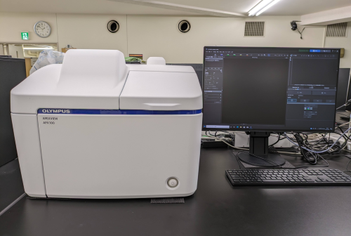
Digital Imaging System
| Model (Manufacturer) |
APX100(EVIDENT) |
| Features and Specifications, etc. |
Overview/Features Since the microscope is housed in a box, a darkroom is not necessary even during fluorescence observation. When the sample is placed on the microscope, it instantly captures a macro image and automatically detects the sample using AI. By changing the sample holder, it is possible to observe 3 slides, 3 dishes with a diameter of 35mm, and multi-well plates. Specifications Inverted Fluorescence Microscope Light source: High color rendering LED, wideband high-power LED Brightfield, fluorescence, phase contrast, gradient contrast Objective Lenses: 4X, 10X, 40X, 60X (Phase Contrast), 20X(Long Working Distance Phase Contrast), 40X (Long Working Distance Phase Contrast) Fluorescence Filters: DAPI,CFP,GFP,YFP,TxRed,CY5.5 (up to 8 cubes can be installed) Cameras: Color CMOS, Monochrome CMOS Z-stack, tiling |
| Location | Central Research Center (University Building 1, Room 317) |
| Remarks | - Please ensure to book this equipment through the reservation system when using it. |
| Equipment Manager Extension Number |
Mizuguchi (Bioimaging Analysis Laboratory) 2645 |
| Usage Fee | 200 yen/hour |
Equipment Photo
Multiplex Spatial analysis system
| Model (Manufacturer) |
CODEX (Akoya) |
| Features and Specifications, etc. |
Overview/Features The tissue microenvironment multiplex spatial analysis system acquires images by linking fluorescence microscopy with immunofluorescence techniques. Antibodies bonded to a unique library of oligonucleotides known as barcodes are used, allowing for the combination of a panel of over 40 CODRX antibodies in a single tissue staining reaction. Images of three different antibodies can be obtained in one cycle by staining tissue with three types of CODEX reporters, each possessing spectrally distinct dyes, and detecting them using antibody barcodes that bind to each respective reporter. By automating this process and repeating it, it becomes possible to detect a wide variety of markers. |
| Location | Central Research Center (Room 317, University Building 1) |
| Remarks | - When using this machine, please ensure to reserve the BZ-X800 simultaneously via the equipment reservation system. |
| Equipment Manager Extension Number |
Mizuguchi (Bioimaging Analysis Laboratory) 2645 |
| Usage Fee | 800 yen/cycle |
Equipment Photo

Virtual Slide Cinematography Equipment
| Model (Manufacturer) |
TOCO24 (Claro) |
| Features and Specifications, etc. |
Overview/Features This device digitizes slide specimens to create high-quality virtual slides. A 10mm square can be captured in about 5 minutes with a 20X lens and about 35 minutes with a 40X lens. It can automatically capture up to 24 slides. Specifications Shooting Lenses: 20X, 40X Maximum number of set specimens: 24 |
| Location | Central Research Center (Room 317, University Building 1) |
| Remarks | - Please ensure to book this equipment through the reservation system when using it. - To take out data, please use a USB or similar device that has been virus-checked by PC for taking out data at the Central Center. |
| Equipment Manager Extension Number |
Mizuguchi (Bioimaging Analysis Laboratory) 2645 |
| Usage Fee | 100 yen/hour |
Equipment Photo

Fluorescence & Bright Field Slide Scanners
| Model (Manufacturer) |
Axioscan7 (Carl Zeiss) |
| Features and Specifications, etc. |
Overview/Features This device digitizes slide specimens (bright field and fluorescence) to create high-quality virtual slides. For a 10mm square in bright field, it can be captured in about 3 minutes with a 20X lens and about 16 minutes with a 40X lens. For fluorescence (four colors), it takes about 24 minutes with a 20X lens and about 100 minutes with a 40X lens. It can automatically capture up to 100 slides. Thick specimens can be captured using Z-stacking. Specifications Shooting Lenses: 20X, 40X Maximum number of set specimens: 100 Fluorescence Channels: DAPI,GFP,TRITC,Cy5 Light Source: LED 385/475/555/630nm |
| Location | Central Research Center (Room 317, University Building 1) |
| Remarks | - Please be sure to apply for eligibility through the equipment reservation system and book your reservation after approval. - To take out data, please use a USB or similar device that has been virus-checked by PC for taking out data at the Central Center. |
| Equipment Manager Extension Number |
Mizuguchi (Bioimaging Analysis Laboratory) 2645 |
| Usage Fee | 200 yen/hour |
Click here for equipment manual
Equipment Photo

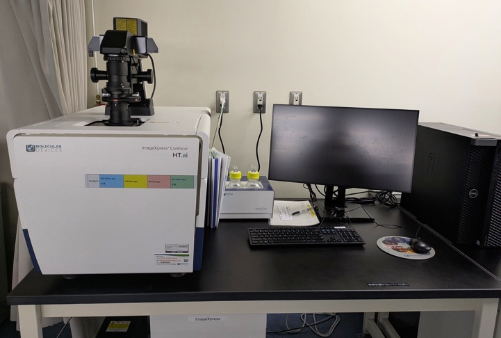
High Content Imaging System
| Model (Manufacturer) |
ImageXpress Confocal HT.ai(Molecular Devices) |
| Features and Specifications, etc. |
Overview/Features - High-throughput image acquisition with short exposure times is possible by utilizing a laser light source with 7 wavelengths corresponding to 8 imaging channels. - Equipped with confocal mode and environmental control unit. The use of a water immersion lens enables deeper observation of thicker samples. Effective combination of MetaXpress software and AI image analysis software simplifies the workflow for advanced phenotyping and 3D image analysis. Samples that can be observed include multi-well plates (1-1536 wells), 35mm dishes, and slides (up to 3 slides can be mounted). Specifications Excitation wavelengths: 405/445/470/520/555/640/730 nm Fluorescence channels: DAPI,CFP,FITC,YFP,TRITC,TxRed,Cy5,Cy7 Acquisition modes: WideField, Confocal Confocal disk: 60㎛ pinhole confocal Objective Lenses: Dry: 4X, 10X, 20X Long Working Distance: 20X, 40X Phase Contrast: 10X, 20X Automatic Immersion: 20X, 40X Time Lapse |
| Location | Central Research Center(Room 317, University Building 1) |
| Remarks | - Please be sure to apply for eligibility through the equipment reservation system and book your reservation after approval. - To take out data, please use a USB or similar device that has been virus-checked by PC for taking out data at the Central Center. |
| Equipment Manager Extension Number |
Watanabe(Oncology Innovation Center)9827 Mizuguchi(Bioimaging Analysis Laboratory)2645 |
| Usage Fee | 400 yen/hour |
Equipment Photo
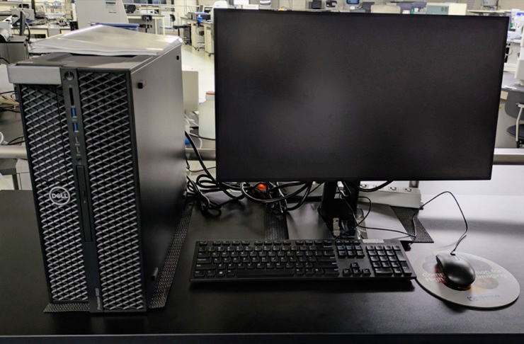
Analysis PC for ImageXpress data
| Model (Manufacturer) |
Precision 5820(Molecular Devices) |
| Features and Specifications, etc. |
 ̄ |
| Location | Central Research Center(Room 317, University Building 1) |
| Remarks | - Please be sure to apply for eligibility through the equipment reservation system and book your reservation after approval. - To take out data, please use a USB or similar device that has been virus-checked by PC for taking out data at the Central Center. |
| Equipment Manager Extension Number |
Watanabe(Oncology Innovation Center)9827 Mizuguchi(Bioimaging Analysis Laboratory)2645 |
| Usage Fee |  ̄ |
Equipment Photo
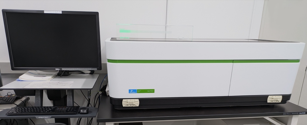
High Content Imaging System
| Model (Manufacturer) |
Opera Phenix (PerkinElmer) |
| Features and Specifications, etc. |
Overview/Features - Automatically captures images of cells labeled with fluorescent markers on microplates (or slides), and quantifies fluorescence and morphological information. - Easy automated image acquisition through an intuitive interface. - Quantification of fluorescence image data, including fluorescence intensity, area, cell count, localization, and positivity rate. - Capable of processing data for a large number of cells. - Suitable for adherent cells, suspension cells, and tissue sections. - Capable of analyzing intracellular localization and subcellular organelle levels. Specifications - Number of cameras: 1 - Laser: 405/488/561/640nm - Bright field: LED brightfield light source - Objective Lenses: 5X, 20X, 40X - A dedicated analysis PC (Second Harmony) is installed separately. |
| Location | Central Research Center Satellite Office(Room 416, University Building 1) |
| Remarks | - Please ensure to book this equipment through the reservation system when using it. - When using the analysis PC (Second Harmony), please make a reservation through the equipment reservation system. |
| Equipment Manager Extension Number |
Mizuguchi (Bioimaging Analysis Laboratory), 2645 |
| Usage Fee | - |
Equipment Photo
Opera Phenix Analysis PC
| Model (Manufacturer) |
Second Harmony (PerkinElmer) |
| Features and Specifications, etc. |
Overview/Features - A dedicated PC for analyzing and exporting data acquired with the Opera Phenix. - Harmony software can be used, similar to the main control PC. |
| Location | Central Research Center Satellite Office(Room 416, University Building 1) |
| Remarks | - Please ensure to book this equipment through the reservation system when using it. - Please ensure to perform a virus check before connecting external hard drives or USB memory sticks. - When using it, you need to start the PC on the Opera Phenix main unit side. |
| Equipment Manager Extension Number |
Mizuguchi (Bioimaging Analysis Laboratory), 2645 |
| Usage Fee | - |
Equipment Photo
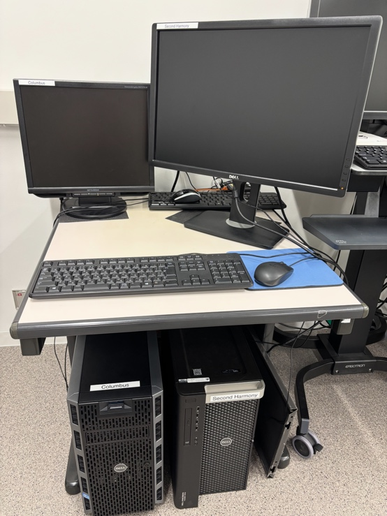
Columbus Image Analysis Processing Software
| Model (Manufacturer) |
Columbus (PerkinElmer) |
| Features and Specifications, etc. |
Overview/Features - Fluorescence Image Analysis Software - Since it runs on a server, you can perform analyses through a browser from your own PC. - Primarily used for analyzing data from the Opera Phenix, but it can also analyze images acquired from other microscopes. - Up to five users can use it simultaneously. |
| Location | Central Research Center Satellite Office(Room 416, University Building 1) |
| Remarks | - No reservation is needed when using it. |
| Equipment Manager Extension Number |
Mizuguchi (Bioimaging Analysis Laboratory), 2645 |
| Usage Fee | - |
Equipment Photo