
Optical Microscope Image Analysis Software
3D/4D Image Analysis Software
| Model (Manufacturer) |
Imaris CF (Bit Plane) |
| Features and Specifications, etc. | Overview/Features Software designed to visualize and analyze 3D/4D image data obtained from confocal and optical microscopes. Software features include measurement of distances between two points in 2D and 3D images, cross-sectional displays at any depth or angle, area and volume measurements, 3D surface rendering, tracing, filament tracing, colocalization, and cell analysis (extracting cells and nuclei as voxels and vesicles as spots, and analyzing relationships between objects). |
| Location | Central Research Center (Room 317, University Building 1) |
| Remarks | - Please ensure to book this equipment through the reservation system when using it. |
| Equipment Manager Extension Number |
Mizuguchi (Bioimaging Analysis Laboratory) 2645 |
| Usage Fee | 100 yen/hour |
Click here for equipment manual
Equipment Photo
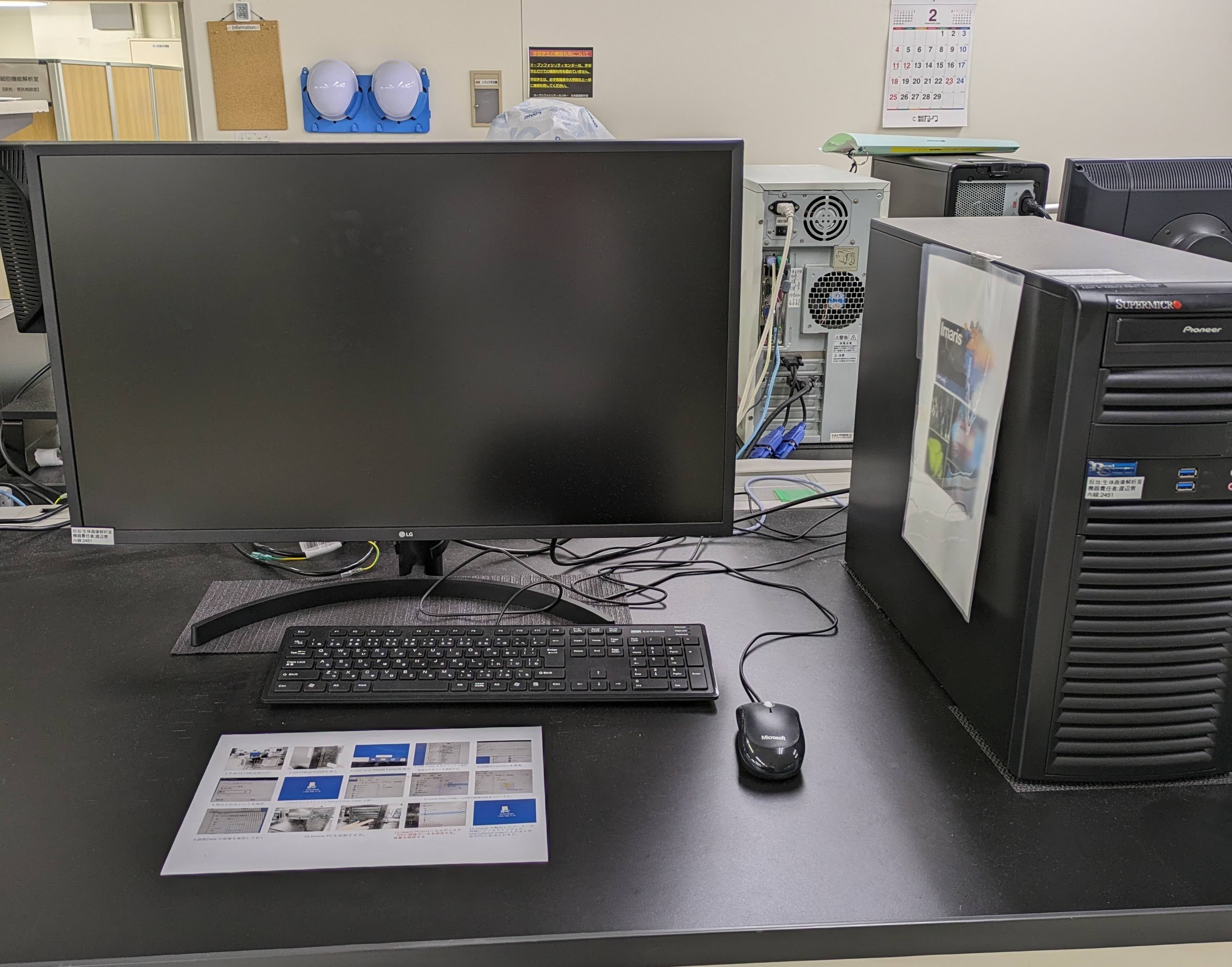
Image Analysis and Processing Software
| Model (Manufacturer) |
BZ-X800,CellSens,ZEN (KEYENCE,EVIDENT,Zeiss) |
| Features and Specifications, etc. |
Overview/Features Software for analysis of images acquired with BZ-9000, BZ-X800, BX-51, APX100, LSM710, LSM980 |
| Location | Central Research Center (Room 317, University Building 1) |
| Remarks | - Please ensure to book this equipment through the reservation system when using it. |
| Equipment Manager Extension Number |
Mizuguchi (Bioimaging Analysis Laboratory) 2645 |
| Usage Fee | - |
Equipment Photo

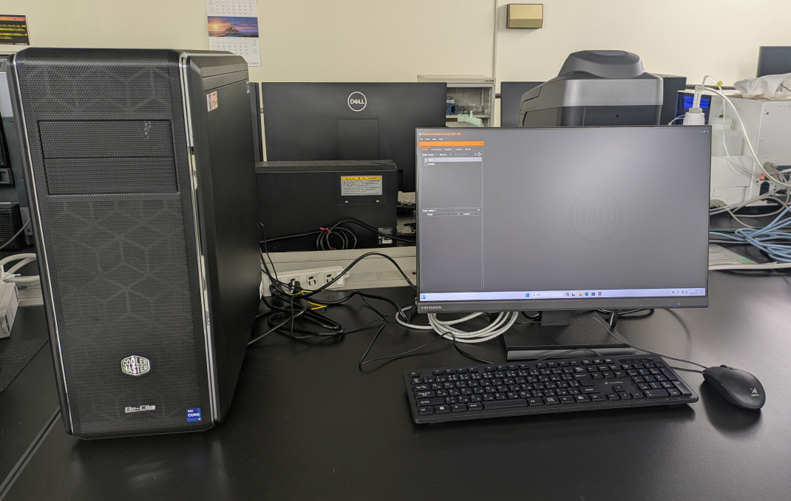
AI Virtual Slide Analysis Software
| Model (Manufacturer) |
HALO(Indica Labs) |
| Features and Specifications, etc. |
Overview/Features - Software for analyzing brightfield images. - Equipped with area quantification, single & multiple IHC, and tissue classification.The Oncology Module features immune cell quantification, serial sections and staining analysis.AI functions are used to achieve more accurate analysis results by allowing them to learn. - Fluorescence images can be quantitatively analyzed for area. |
| Location | Central Research Center(Room 317, University Building 1) |
| Remarks | - This equipment was introduced under the Next Generation Cancer Professional Training Plan Project. - Use by Outside the University persons is limited to members of the Next Generation Cancer Professional Development Plan Project Cooperating Universities. - Please ensure to book this equipment through the reservation system when using it. |
| Equipment Manager Extension Number |
Mizuguchi (Bioimaging Analysis Laboratory) 2645 |
| Usage Fee | ― |
Equipment Photo