

Spectrometer
Ultra-Trace Spectrophotometer
| Model (Manufacturer) |
NanoDrop One (ThermoFisher) |
| Features and Specifications, etc. | This device cannot be used for turbidity measurements of microorganisms (such as E. coli). (Microorganisms, infectious samples, etc., cannot be brought into OFC facilities.) DNA, RNA, and protein samples of 1-2 µL can be accurately quantified in seconds. Dynamic range: 2.0-27,500 ng/µL - Rapid measurement: Automatic measurement starts when the arm is lowered, allowing for quick and easy quantification of nucleic acids and proteins using pre-programmed applications. - Accurate measurements up to 27,500 ng/μL (dsDNA) are possible with automatic path length adjustment. - Samples are directly dropped onto the pedestal using a pipette, eliminating the need for preprocessing and consumables such as measurement plates. 【Specifications】 Minimum sample volume: 1 µL Detection limit for dsDNA pedestal: 2.0 ng/µL Detection limit for BSA (IgG) pedestal: 0.06 (0.03) mg/mLDetection limit for dsDNA pe-destal: 27,500 ng/μL Detection limit for BSA (IgG) pedestal: 820 (400) mg/mL Time from measurement to data processing: 8 seconds Wavelength range: 190-850 nm Absorbance range (equivalent to 10 mm path length) pedestal: 0-550 A Installed in September 2020 |
| Location | Central Research Center (Room 317, University Building 1) |
| Remarks | - |
| Equipment Manager Extension Number |
Maeda (Omics Analysis Laboratory) 9814 |
| Usage Fee | 100 yen per sample |
Equipment Photo

Absorption Microplate Reader
| Model (Manufacturer) |
Benchmark (BioRad) |
| Features and Specifications, etc. | It measures the optical properties of samples (mainly liquids) placed in a microplate. ELISA (measurement of color development by absorbance) Enzyme activity (such as chromogenic substrates) Quantification of proteins, nucleic acids, etc. (absorbance) 【Specifications】 Measurement wavelengths: 405, 450, 490, 550, 595, 655 nm Measurement plate: 96-well microplate |
| Location | Central Research Center (Room 317, University Building 1) |
| Remarks | - |
| Equipment Manager Extension Number |
Maeda (Omics Analysis Laboratory) 9814 |
| Usage Fee | - |
Equipment Photo

Absorption Microplate Reader
| Model (Manufacturer) |
SUNRISE Rainbow (Fujifilm Wako) |
| Features and Specifications, etc. | It measures the optical properties of samples (mainly liquids) placed in a microplate. Equipped with a rainbow filter that can select wavelengths from 400 nm to 700 nm in 1 nm steps. The measurement wavelength range when using standard filters is from 340 nm to 750 nm. In addition to endpoint measurements, kinetic measurements and spectral measurements are possible. 【Specifications】 Wavelength variable: 400 to 700 nm (when using the rainbow filter) Wavelength range: 340 to 750 nm Measurement range: 0 to 4.0 OD (400–750 nm) Measurement time for 96-well plate: 8 seconds for 1 wavelength, 16 seconds for 2 wavelengths |
| Location | Maeda (Omics Analysis Laboratory) 9814 |
| Remarks | - |
| Equipment Manager Extension Number |
Maeda (Omics Analysis Laboratory) 9814 |
| Usage Fee | - |
Equipment Photo

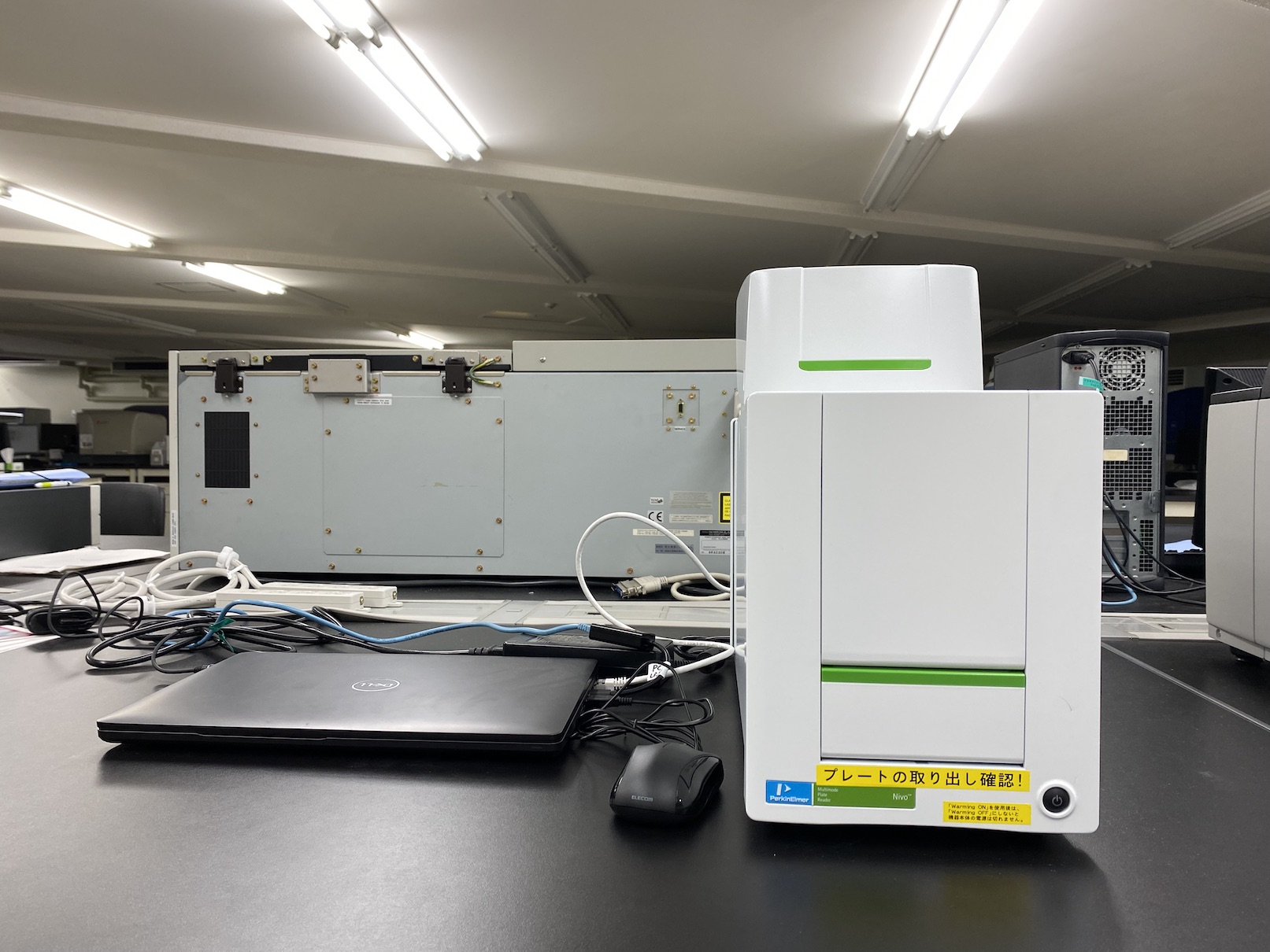
Absorbance, Luminescence, and Fluorescence Microplate Reader
| Model (Manufacturer) |
Nivo S Dispencer (PerkinElmer) |
| Features and Specifications, etc. | A microplate reader for measuring absorbance, luminescence, fluorescence intensity, time-resolved fluorescence, and fluorescence polarization. 【Specifications】 - Absorbance measurement (spectrometer) : 230 nm to 1000 nm (resolution: 2 nm, 5 nm, 10 nm) - Measurements from both the top and bottom of the plate (excluding absorbance) - Temperature control up to 65°C - Equipped with dispensers (dual injectors) : Automatic dispensing function - Model capable of measuring time-resolved fluorescence and fluorescence polarization - For time-resolved fluorescence and fluorescence polarization measurements, corresponding dichroic mirrors and filters for the application must be prepared. - Data analysis software: My Assays 【Fluorescence Filters】 - Equipped with fluorescence rainbow filter 355/40 nm, 435/20 nm, 460/30 nm, 495/20 nm, 540/30 nm, 580/20 nm, 625/30 nm, 640/30 nm, 685/30 nm 【Filters】 - 405/10 nm (standard included) - 480/30 nm (standard included) - 530/30 nm (standard included) x 2 - 700 nm SP (standard included) : For luminescence measurement - 740/40 nm (addition) - 780 nm long-pass (addition) 【Dichroic mirrors】 - BS50/50 (standard included) - D500: For fluorescence polarization - D660 (addition) - D770 (addition) Installed in February 2021 |
| Location | Central Research Center (Room 317, University Building 1) |
| Remarks | - Please ensure to book this equipment through the reservation system when using it. - When using the equipment, please be sure to fill out the logbook provided with it. - Please read the usage notes carefully before using the logbook. - If the connection between the device and the PC is lost, disconnect the "PC LAN cable" from the PC and proceed with the measurement. |
| Equipment Manager Extension Number |
Tsukamoto (Laboratory for Proteins and Genes) 9928 |
| Usage Fee | 100 yen/time |
Equipment Photo
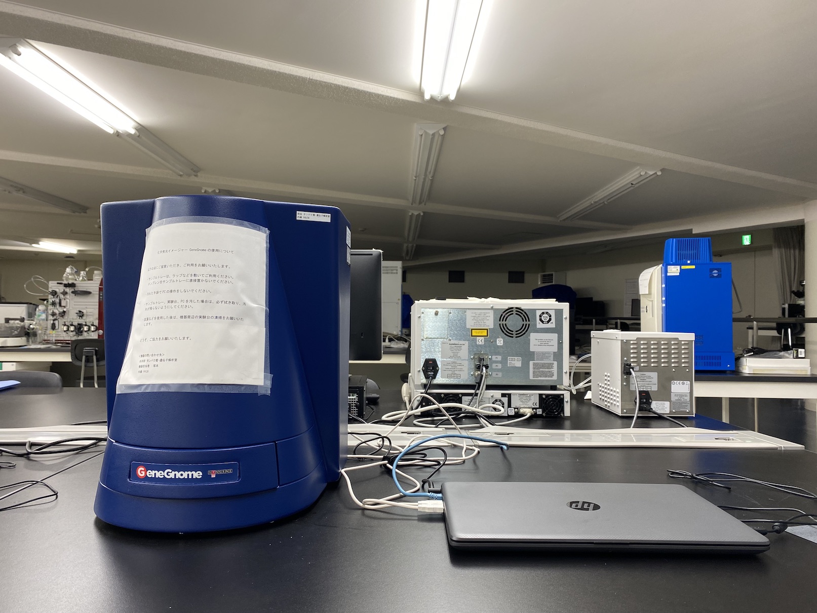
Chemiluminescent Imager
| Model (Manufacturer) |
GeneGnome (SYGENE) |
| Features and Specifications, etc. | A device for chemiluminescence detection (chemi-luminescence imaging detection), imaging, and image analysis. Installed in January 2022 【Specifications】 - Resolution: 4 M pixels (16 M pixels) - Grayscale: 65,536 shades of black and white - Camera: Cooled digital CCD (16-bit) - Lens: F0.95 fixed lens - White light source: Built-in white LED - Resolutions: 21, 42, 84, 169, 337 µm - Control software: GeneSys (image capturing software) - Image analysis software: GeneTools (automatic analysis software) Installed in November 2021 |
| Location | Central Research Center (Room 317, University Building 1) |
| Remarks | - Please ensure to book this equipment through the reservation system when using it. - When using the equipment, please be sure to fill out the logbook provided with it. - Please read and follow the instructions displayed on the device before use. |
| Equipment Manager Extension Number |
Tsukamoto (Laboratory for Proteins and Genes) 9928 |
| Usage Fee | 100 yen/time |
Equipment Photo
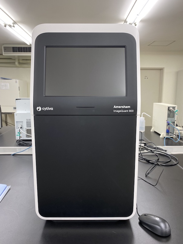
Chemiluminescent Imager
| Model (Manufacturer) |
ImageQuant 800(Cytiva) |
| Features and Specifications, etc. | A device for chemiluminescence detection (chemi-luminescence imaging detection), imaging of CBB or silver-stained gels, and image analysis. 【Specifications】 Model: ImageQuant 800 System - Grayscale: 65,536 levels (16-bit) - Camera: Cooled digital CCD (-25°C) - CCD type: 8.3 megapixel high-resolution Peltier cooled CCD - Lens: F0.74 (Fujifilm) - White light source: epi-illumination LED - Detection area (max.) : upper tray guide 80 x 110 mm, lower tray guide 160 x 220 mm - Dynamic range: 4.8 orders - Exposure time: 1/10 second to 10 hours. Resolution 1/10 second - Image analysis software: ImageQuantTL Installed in June 2022 |
| Location | Central Research Center (Room 317, University Building 1) |
| Remarks | - Do not place samples directly on the black tray and white insert. Wrap the sample in plastic wrap or similar material. - Please handle with care, as scratches or dirt on the black tray can affect image capture. - Please ensure to book this equipment through the reservation system when using it. - When using the equipment, please be sure to fill out the logbook provided with it. - Please read and follow the instructions displayed on the device before use. |
| Equipment Manager Extension Number |
Tsukamoto (Laboratory for Proteins and Genes) 9928 |
| Usage Fee | 100 yen/time |
Equipment Photo
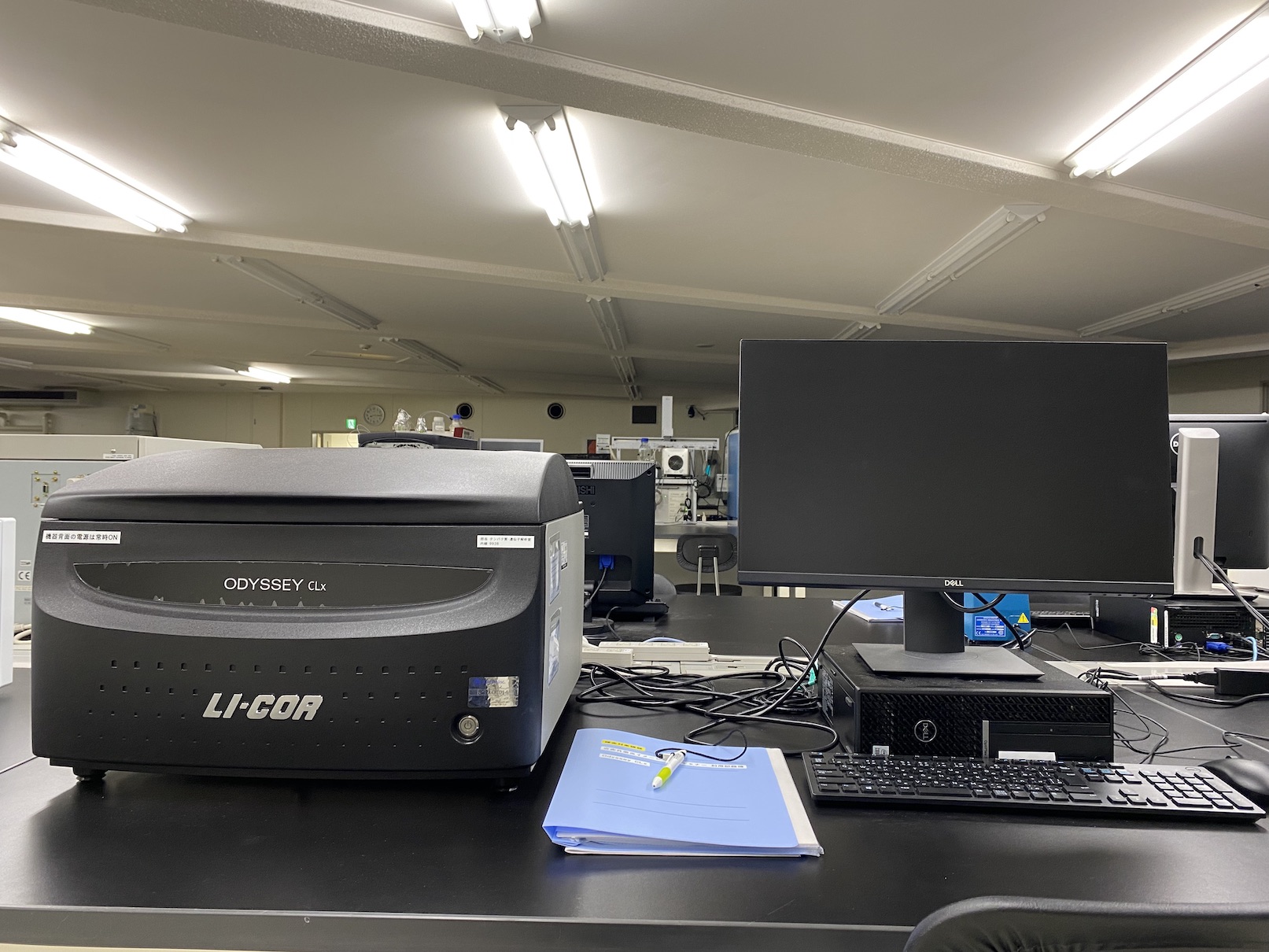
Near Infrared Fluorescence Imaging Scanner
| Model (Manufacturer) |
Odyssey CLx (LI-COR) |
| Features and Specifications, etc. | A device used for protein expression analysis through fluorescent Western blotting and In-Cell Western assays. 【Features】 - Fluorescent Western blotting is a suitable method for quantitative Western blotting, providing highly reliable data with better reproducibility and linearity compared to chemiluminescence methods. - The In-Cell Western assay method allows for high-throughput protein expression analysis directly in cultured cells without the need for protein extraction or other preparatory steps. This experimental method is commonly used in fields such as drug discovery, vaccine development, and microbiome research. - It can also be used for applications such as gel shift assays (EMSA), protein arrays, and organ imaging. 【Specifications】 - Method: Point scanner - Imaging area: 25 x 25 cm - Pump light sources: 685 nm semiconductor laser, 785 nm semiconductor laser - Detection channels: 700: Ex 685 nm / Em 710-730 nm, 800: Ex 785 nm / Em 812-832 nm - Detector: Silicon avalanche photodiodes - Resolution: 21, 42, 84, 169, 337 µm - Dynamic range: 】 6 digits - Focus: 0 to 4 mm - Image analysis software: Empiria Studio (can also be used with "Protein and Gene Analysis PC") Installed in February 2021 |
| Location | Central Research Center (Room 317, University Building 1) |
| Remarks | After use, please clean the glass surface following these steps: 1. Drop a small amount of Milli-Q water onto the glass surface and thoroughly wipe it off with a pro-wipe or lens paper. 2. Apply a small amount of 70% ethanol to the glass surface and wipe it off thoroughly. 3. Use an air duster to blow away any dust. - Please ensure to book this equipment through the reservation system when using it. - When using the equipment, please be sure to fill out the logbook provided with it. - Please read the usage notes carefully before using the logbook. - If you need to use the image analysis software (Empiria Studio) for an extended period, please perform the analysis on the " Proteins and Genes Analysis PC." Also, please make a reservation for a PC. |
| Equipment Manager Extension Number |
Tsukamoto (Laboratory for Proteins and Genes) 9928 |
| Usage Fee | 100 yen/time |
Equipment Photo
Fluorescence / Luminescence Plate imager
| Model (Manufacturer) |
FDSS/μCELL(Hamamatsu Photonics) |
| Features and Specifications, etc. | - Suitable for fluorescence/luminescence analysis - Simultaneous addition and reading in 96 wells/384wells - Enables a wide range of measurements with excitation light sources of various wabelengthes - Long life, high power LED excitation light source - Suitable for FRET or BRET by changing wavelength - High speed data capture of 5ms maximum(Optopnal) 【Specifications】 1 Head 1 Plate Compatible Dispensing Head: 96-channel chip type, 384-channel chip type Cleaning Tank: For 96-chip, for 384-chip Chip Removal and Attachment Device Heater Unit Excitation light source unit (B, G): Excitation LED 480nm, 530nm, fluorescent filter 530nm, 590nm Fluorescence/emission sensor: EMCCD camera 【Available indicator examples】 For Ca2+: Fluo4 (Fluo8), Cal520, CalBryte series, Cal590, etc. For membrane potential: FluoVolt, FMP, ScreenQuest Membrane Potential Assay Kit, etc. |
| Location | University Building 1, Room 315 |
| Remarks | - If you wish to use this equipment, please contact the responsible personnel. (We will eventually transition to a equipment reservation system) - When using the equipment, please be sure to fill out the logbook provided with it. |
| Equipment Manager Extension Number |
Yukiko Moriguchi,Daisuke Tsuboi 9576 |
| Usage Fee | - |