

Optical Microscope Specimen Preparation Device
Contents on page
- Ultrasonic Automatic Embedding Device
- Paraffin Embedding Device
- Paraffin Embedding Center
- Cryomicrotome
- Cryomicrotome
- Sliding Microtome Unit 1
- Sliding Microtome Unit 2
- Rotary Microtome
- Staining Series
- Pressure Cooker and Hot Plate for Antigen Retrieval
- Paraffin Section Dryer
- Microwave Processing Device
- Instrument Dryer
- Incubator
- [Dedicated Consignment Machine] Paraffin Embedding Device
- [Dedicated Consignment Machine] Paraffin Embedding Block Preparation Device
- [Dedicated Consignment Machine] Sliding Microtome
- [Dedicated Consignment Machine] Fully Automatic Immunostaining Device
Ultrasonic Automatic Embedding Device
| Model (Manufacturer) |
Histra-QS (JOKOH) |
| Features and Specifications, etc. | Overview/Features - A specimen processing device that automatically performs dehydration, degreasing, and paraffin infiltration of specimens. - Intuitive operation is possible through a touchscreen display. - Processes from dehydration to paraffin infiltration in as little as 58 minutes. - Capable of processing up to 30 cassettes simultaneously. - It is a compact, desktop-sized device that uses a small amount of reagent, reducing running costs. - Ultrasonic treatment enhances the uniform penetration of reagents and improves various staining properties. Specifications - Treatment principle: Rapid treatment using ultrasound - Number of specimens processed simultaneously: 30 cassettes - Temperature range: 40°C to 80°C (in chemical tanks, paraffin tanks, and retorts) - Agitation method: ultrasonic vibration - Number of stored programs: 6 - Display: Touch panel type display - Exhaust gas treatment: Deodorizing filter using activated carbon - Ultrasonic oscillation frequency: 40 kHz |
| Location | Bioimaging Analysis Laboratory (Pathology Specimen Preparation Room, Room 114, University Building 1) |
| Remarks | - This device can only be used by individuals who have completed viewing the equipment usage tutorial video and have registered their names in the equipment users log. - Please ensure to book this equipment through the reservation system when using it. - When using the equipment, please be sure to fill out the logbook provided with it. |
| Equipment Manager Extension Number |
Tominaga (Bioimaging Analysis Laboratory) 2320 |
| Usage Fee | 400 yen/run |
Equipment Photo

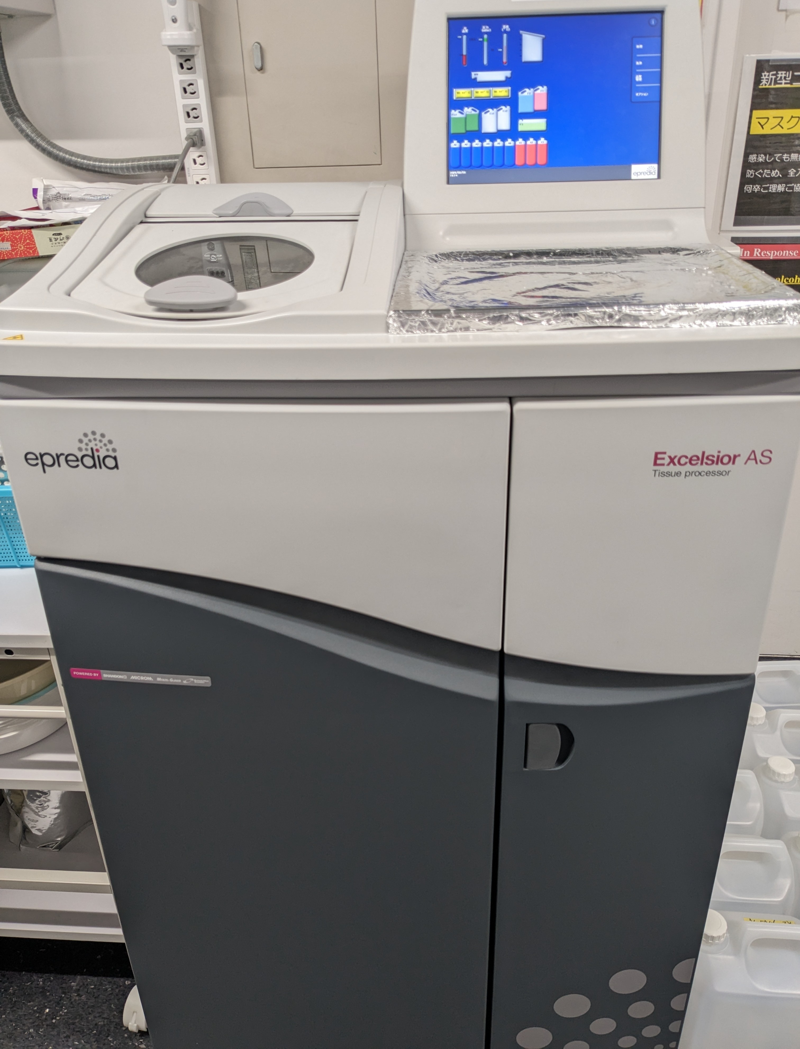
Paraffin Embedding Device
| Model (Manufacturer) |
Excelsior AS (PHC) |
| Features and Specifications, etc. | Overview/Features - A specimen processing device that automatically performs specimen dehydration, degreasing, and paraffin penetration. - Up to 300 cassettes can be processed simultaneously in a single operation. - There are 14 processing steps, with 11 chemical solution steps and 3 paraffin steps performed automatically. - The cassette basket rotates gently within the circular processing layer, allowing for effective circulation of reagents. - The alcohol density system optimizes reagent exchange timing, helping to reduce costs. - The downdraft ventilation system automatically activates, neutralizing harmful gases through a filter. Specifications - Maximum simultaneous sample processing: 300 samples - Exhaust gas treatment: Activated carbon filter, condenser - Mixing method: Cassette basket rotation - Display: Touchscreen |
| Location | Bioimaging Analysis Laboratory (Pathology Specimen Preparation Room, Room 114, University Building 1) |
| Remarks | - Please ensure to book this equipment through the reservation system when using it. - When using the equipment, please be sure to fill out the logbook provided with it. - Basically, please make a reservation for a 1:00-3:00 p.m. set time and a 9:00-10:00 a.m. end time the next day. |
| Equipment Manager Extension Number |
Tominaga (Bioimaging Analysis Laboratory) 2320 |
| Usage Fee | 1,500 yen/run |
Equipment Photo
Paraffin Embedding Center
| Model (Manufacturer) |
Tissue-Tek TEC-P (SAKURA) |
| Features and Specifications, etc. | Overview/Features - This specimen processing device is used for embedding tissues that have completed paraffin infiltration, as part of the series of processes involved in tissue specimen preparation. - This device features a cooling section to facilitate the positioning of tissue slices within the embedding block, as well as functions to hold and dispense molten paraffin for forming the embedding block. Specifications - Temperature control: 50°C to 75°C (paraffin tank, heating tank, hot plate) 15°C or lower (cold spot) |
| Location | Bioimaging Analysis Laboratory (Pathology Specimen Preparation Room, Room 114, University Building 1) |
| Remarks | - Please ensure to book this equipment through the reservation system when using it. - When using the equipment, please be sure to fill out the logbook provided with it. |
| Equipment Manager Extension Number |
Tominaga (Bioimaging Analysis Laboratory) 2320 |
| Usage Fee | 100 yen/hour, 30 yen/block |
Equipment Photo

Cryomicrotome
| Model (Manufacturer) |
CM1950 (Leica) |
| Features and Specifications, etc. | Overview/Features - Equipment for creating frozen pathology specimens. - To prevent infection during and after operations, the equipment maintains a clean state at all times, equipped with a UVC freezing chamber disinfection system and AgProtect antibacterial nano-silver coating. - The device can be comfortably operated using single-function keys and an easy-to-read LED display. Specifications - Temperature range: Sample table (-10°C to -50°C) Library (0°C to -35°C) - Minimum temperature for quick-freezing station: -42°C - Number of quick-freezing stations: 15 - Thin slice thickness setting range: 1 to 100 μm - Infection prevention function: UVC disinfection 30 min. |
| Location | Bioimaging Analysis Laboratory (Pathology Specimen Preparation Room, Room 114, University Building 1) |
| Remarks | - This device can only be used by individuals who have completed viewing the equipment usage tutorial video and have registered their names in the equipment users log. - Please ensure to book this equipment through the reservation system when using it. - When using the equipment, please be sure to fill out the logbook provided with it. - Please prepare blades for thin slicing in each laboratory. |
| Equipment Manager Extension Number |
Tominaga (Bioimaging Analysis Laboratory) 2320 |
| Usage Fee | 100 yen/hour |
Equipment Photo

Cryomicrotome
| Model (Manufacturer) |
Tissue-Tek Polar (SAKURA) |
| Features and Specifications, etc. | Overview/Features - Equipment for creating frozen pathology specimens. - To prevent infection during and after operations, the equipment features an ozone treatment function and a vacuum feature, along with a draft generator that reduces the spread of contaminated air outside the cabinet. - Thanks to its color touchscreen display, intuitive operation is possible. Specifications - Temperature range: Sample table (-10°C to -50°C) Library (-10°C to -35°C) - Cryobar temperature: -40°C or lower - Number of cryobar samples: 12 (square) or 15 (round) - Thin slice thickness setting range: 1 to 99 μm - Infection prevention function: 75 minutes of ozone treatment (including 15 minutes of ozone treatment) - Display: 6.2" VGA TFT color LCD display |
| Location | Bioimaging Analysis Laboratory (Pathology Specimen Preparation Room, Room 114, University Building 1) |
| Remarks | - This device can only be used by individuals who have completed viewing the equipment usage tutorial video and have registered their names in the equipment users log. - Please ensure to book this equipment through the reservation system when using it. - When using the equipment, please be sure to fill out the logbook provided with it. - Please prepare blades for thin slicing in each laboratory. |
| Equipment Manager Extension Number |
Tominaga (Bioimaging Analysis Laboratory) 2320 |
| Usage Fee | 100 yen/hour |
Equipment Photo

Sliding Microtome Unit 1
| Model (Manufacturer) |
REM-710 (Yamato Kohki) |
| Features and Specifications, etc. | Overview/Features - Equipment used to create pathology specimens from embedding blocks encased in paraffin or similar materials. - A microtome that combines the precision of digital technology with the tactile feel of analog controls, offering superior operability. - When the cutting edge returns backward, the retraction function lowers the sample to avoid contact with the back surface of the blade. It also includes many features such as the auto-reverse function, which automatically corrects when changing knife holders or thickness settings. |
| Location | Bioimaging Analysis Laboratory (Pathology Specimen Preparation Room, Room 114, University Building 1) |
| Remarks | - Please ensure to book this equipment through the reservation system when using it. - When using the equipment, please be sure to fill out the logbook provided with it. |
| Equipment Manager Extension Number |
Tominaga (Bioimaging Analysis Laboratory) 2320 |
| Usage Fee | - |
Equipment Photo

Sliding Microtome Unit 2
| Model (Manufacturer) |
REM-710 (Yamato Kohki) |
| Features and Specifications, etc. | Overview/Features - Equipment used to create pathology specimens from embedding blocks encased in paraffin or similar materials. - A microtome that combines the precision of digital technology with the tactile feel of analog controls, offering superior operability. - When the cutting edge returns backward, the retraction function lowers the sample to avoid contact with the back surface of the blade. It also includes many features such as the auto-reverse function, which automatically corrects when changing knife holders or thickness settings. |
| Location | Bioimaging Analysis Laboratory (Pathology Specimen Preparation Room, Room 114, University Building 1) |
| Remarks | - Please ensure to book this equipment through the reservation system when using it. - When using the equipment, please be sure to fill out the logbook provided with it. |
| Equipment Manager Extension Number |
Tominaga (Bioimaging Analysis Laboratory) 2320 |
| Usage Fee | - |
Equipment Photo

Rotary Microtome
| Model (Manufacturer) |
RM2125RT (Leica) |
| Features and Specifications, etc. | Overview/Features - Equipment used to create pathology specimens from embedding blocks encased in paraffin or similar materials. - With a two-stage trimming process (rough trimming at 50μm, and fine trimming at 10μm), it is possible to quickly prepare tissue sections. |
| Location | Bioimaging Analysis Laboratory (Pathology Specimen Preparation Room, Room 114, University Building 1) |
| Remarks | - Please ensure to book this equipment through the reservation system when using it. - When using the equipment, please be sure to fill out the logbook provided with it. |
| Equipment Manager Extension Number |
Tominaga (Bioimaging Analysis Laboratory) 2320 |
| Usage Fee | - |
Equipment Photo

Staining Series
| Model (Manufacturer) |
- |
| Features and Specifications, etc. | Overview/Features - It is possible to perform deparaffinization, hydration, staining, dehydration, and clearing. |
| Location | Bioimaging Analysis Laboratory (Pathology Specimen Preparation Room, Room 114, University Building 1) |
| Remarks | - Please ensure to book this equipment through the reservation system when using it. - When using the equipment, please be sure to fill out the logbook provided with it. - Please prepare the reagents used for staining in each laboratory. |
| Equipment Manager Extension Number |
Tominaga (Bioimaging Analysis Laboratory) 2320 |
| Usage Fee | - |
Equipment Photo

Pressure Cooker and Hot Plate for Antigen Retrieval
| Model (Manufacturer) |
CLPSO+ (T-fal) KZ-PH5P (Panasonic) |
| Features and Specifications, etc. | Overview/Features - Pressure cookers are used for antigen retrieval during immunostaining. - Hot plates are used for drying thin tissue sections and during the drying process for embedding. |
| Location | Bioimaging Analysis Laboratory (Pathology Specimen Preparation Room, Room 114, University Building 1) |
| Remarks | - Please ensure to book this equipment through the reservation system when using it. - When using the equipment, please be sure to fill out the logbook provided with it. |
| Equipment Manager Extension Number |
Tominaga (Bioimaging Analysis Laboratory) 2320 |
| Usage Fee | - |
Equipment Photo

Paraffin Section Dryer
| Model (Manufacturer) |
SIB-35 (SANSYO) |
| Features and Specifications, etc. | Overview/Features - This device is used for drying paraffin sections prepared with a microtome. - The set temperature is 60°C. Specifications - Heating method: Natural convection - Heater: Silicon cord heater - Temperature control range: room temperature +5°C to 60°C - Internal volume: 35L |
| Location | Bioimaging Analysis Laboratory (Pathology Specimen Preparation Room, Room 114, University Building 1) |
| Remarks | - When using the equipment, please be sure to fill out the logbook provided with it. |
| Equipment Manager Extension Number |
Tominaga (Bioimaging Analysis Laboratory) 2320 |
| Usage Fee | - |
Equipment Photo

Microwave Processing Device
| Model (Manufacturer) |
MI-77 (Azumaya Medical Instruments) |
| Features and Specifications, etc. | Overview/Features - Equipment used for immunohistochemical staining reactions, as well as for hybridization processing in FISH (Fluorescence In Situ Hybridization) and in situ hybridization. - It is capable of controlling microwave output, measuring precise temperatures, and irradiating microwaves while monitoring temperature accurately. |
| Location | Bioimaging Analysis Laboratory (Pathology Specimen Preparation Room, Room 114, University Building 1) |
| Remarks | - Please ensure to book this equipment through the reservation system when using it. - When using the equipment, please be sure to fill out the logbook provided with it. |
| Equipment Manager Extension Number |
Tominaga (Bioimaging Analysis Laboratory) 2320 |
| Usage Fee | - |
Equipment Photo

Instrument Dryer
| Model (Manufacturer) |
- (AS ONE) |
| Features and Specifications, etc. | Overview/Features - Equipment for drying used tools such as staining baskets and metal embedding trays. |
| Location | Bioimaging Analysis Laboratory (Pathology Specimen Preparation Room, Room 114, University Building 1) |
| Remarks | - When using the equipment, please be sure to fill out the logbook provided with it. |
| Equipment Manager Extension Number |
Tominaga (Bioimaging Analysis Laboratory) 2320 |
| Usage Fee | - |
Equipment Photo

Incubator
| Model (Manufacturer) |
SIB-35 (SANSYO) |
| Features and Specifications, etc. | Overview/Features - Equipment used for enzyme reactions in immunostaining and for special staining techniques that require temperature control. - The set temperature is 37°C to 60°C. Specifications - Heating method: Natural convection - Heater: Silicon cord heater - Temperature control range: room temperature +5°C to 60°C - Internal volume: 35L |
| Location | Bioimaging Analysis Laboratory (Pathology Specimen Preparation Room, Room 114, University Building 1) |
| Remarks | - Please ensure to book this equipment through the reservation system when using it. - When using the equipment, please be sure to fill out the logbook provided with it. |
| Equipment Manager Extension Number |
Tominaga (Bioimaging Analysis Laboratory) 2320 |
| Usage Fee | - |
Equipment Photo

[Dedicated Consignment Machine]
Paraffin Embedding Device
| Model (Manufacturer) |
VIP5Jr (Sakura Finetek Japan) |
| Features and Specifications, etc. | Overview/Features - An automatic specimen preprocessing device (fixation and paraffin embedding system) used in histological research and examinations in pathology, anatomy, and clinical pathology, which automates the processing of samples (tissue specimens) required for specimen preparation at each stage. Specifications - Number of cassettes processed per batch: 150 cassettes (when Unicassette is used) - Number of treatment processes: 14 (10 chemicals, 4 paraffin) |
| Location | Bioimaging Analysis Laboratory (Pathology Specimen Preparation Room, Room 114, University Building 1) |
| Remarks | - This equipment is used exclusively for contracted services in the production of light microscopy specimens in the bioimaging analysis laboratory. Therefore, it is not available for use by the general public. |
| Equipment Manager Extension Number |
Tominaga (Bioimaging Analysis Laboratory) 2320 |
| Usage Fee | - |
Equipment Photo

[Dedicated Consignment Machine]
Paraffin Embedding Block Preparation Device
| Model (Manufacturer) |
TEC-P-S-J0 (Sakura Finetek Japan) |
| Features and Specifications, etc. | Overview/Features - This is a specimen preprocessing device (paraffin embedding block creation device) used in the production of tissue samples in histological research and examinations in pathology, anatomy, and clinical pathology. It is used specifically for embedding tissues that have undergone paraffin infiltration, to perform paraffin embedding. - Paraffin embedding blocks are created for the purpose of slicing tissue specimens thinly using a medical microtome. The process involves placing tissue sections and melted paraffin into an embedding mold (a mold used to create the block) and then solidifying them by cooling. - This device is equipped with a cooling section to facilitate the positioning of tissue sections within the embedding block, as well as features for holding and dispensing melted paraffin necessary for forming the embedding block. Specifications - Temperature control: 50°C to 75°C (paraffin tank, heating tank, hot plate) 15°C or lower (cold spot) |
| Location | Bioimaging Analysis Laboratory (Pathology Specimen Preparation Room, Room 114, University Building 1 ) |
| Remarks | - This equipment is used exclusively for contracted services in the production of light microscopy specimens in the bioimaging analysis laboratory. Therefore, it is not available for use by the general public. |
| Equipment Manager Extension Number |
Tominaga (Bioimaging Analysis Laboratory) 2320 |
| Usage Fee | - |
Equipment Photo

[Dedicated Consignment Machine]
Sliding Microtome
| Model (Manufacturer) |
REM-710 (Yamato Kohki) |
| Features and Specifications, etc. | Overview/Features - Equipment used to create pathology specimens from embedding blocks encased in paraffin or similar materials. - A microtome that combines the precision of digital technology with the tactile feel of analog controls, offering superior operability. - When the cutting edge returns backward, the retraction function lowers the sample to avoid contact with the back surface of the blade. It also includes many features such as the auto-reverse function, which automatically corrects when changing knife holders or thickness settings. Specifications - Vertical movement distance: 40mm - Sliding method: Crossed roller bearing sliding |
| Location | Bioimaging Analysis Laboratory (Pathology Specimen Preparation Room, Room 114, University Building 1) |
| Remarks | - This equipment is used exclusively for contracted services in the production of light microscopy specimens in the bioimaging analysis laboratory. Therefore, it is not available for use by the general public. |
| Equipment Manager Extension Number |
Tominaga (Bioimaging Analysis Laboratory) 2320 |
| Usage Fee | - |
Equipment Photo

[Dedicated Consignment Machine]
Fully Automatic Immunostaining Device
| Model (Manufacturer) |
BOND RX (Leica Biosystems) |
| Features and Specifications, etc. | Overview/Features - This device is a fully automatic immunostaining machine for research purposes, capable of automatically performing various tests such as immunohistochemical staining and in situ hybridization. Specifications - Slide capacity: 30 slides - Slide width: 24.64 to 26.0 mm - Slide length: 74.9 to 76.0 mm - Slide thickness: 0.8 to 1.3 mm - Number of reagent containers: 36 - Chemical compatibility: All Bond reagents |
| Location | Bioimaging Analysis Laboratory (Pathology Specimen Preparation Room, Room 114, University Building 1 ) |
| Remarks | - This equipment is used exclusively for contracted services in the production of light microscopy specimens in the bioimaging analysis laboratory. Therefore, it is not available for use by the general public. |
| Equipment Manager Extension Number |
Tominaga (Bioimaging Analysis Laboratory) 2320 |
| Usage Fee | - |
Equipment Photo